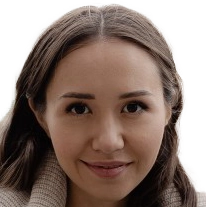
Research Group Nassir Navab
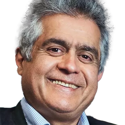
Nassir Navab
holds the Chair of Computer Aided Medical Procedures & Augmented Reality at TU Munich.
His research focuses on computer-aided medical procedures and augmented reality. The work involves developing technologies to improve the quality of medical intervention and bridges the gap between medicine and computer science.
Team members @MCML
PostDocs
PhD Students
Recent News @MCML
Publications @MCML
2026
[165]

A. Topaloglu • K. Li • M. Niemeyer • N. Navab • A. M. Tekalp • F. Tombari
OracleGS: Grounding Generative Priors for Sparse-View Gaussian Splatting.
WACV 2026 - IEEE/CVF Winter Conference on Applications of Computer Vision. Tucson, AZ, USA, Mar 06-10, 2026. To be published. Preprint available. arXiv
OracleGS: Grounding Generative Priors for Sparse-View Gaussian Splatting.
WACV 2026 - IEEE/CVF Winter Conference on Applications of Computer Vision. Tucson, AZ, USA, Mar 06-10, 2026. To be published. Preprint available. arXiv
[164]
F. Li • Y. Bi • T. Song • Z. Jiang • N. Navab
Ultrasound-Guided Real-Time Spinal Motion Visualization for Spinal Instability Assessment.
Preprint (Feb. 2026). arXiv
Ultrasound-Guided Real-Time Spinal Motion Visualization for Spinal Instability Assessment.
Preprint (Feb. 2026). arXiv
[163]
T. Song • F. Li • F. Pabst • M.-A. Gafencu • Y. Bi • U. Eck • N. Navab
Comparative Study of Ultrasound Shape Completion and CBCT-Based AR Workflows for Spinal Needle Interventions.
Preprint (Feb. 2026). arXiv
Comparative Study of Ultrasound Shape Completion and CBCT-Based AR Workflows for Spinal Needle Interventions.
Preprint (Feb. 2026). arXiv
[162]

M. Wei • K. Yuan • S. Li • Y. Zhou • L. Bai • N. Navab • H. Ren • H. J. Lee • T. Vercauteren • N. Padoy
Where It Moves, It Matters: Referring Surgical Instrument Segmentation via Motion.
AAAI 2026 - 40th Conference on Artificial Intelligence. Singapore, Jan 20-27, 2026. To be published. Preprint available. arXiv
Where It Moves, It Matters: Referring Surgical Instrument Segmentation via Motion.
AAAI 2026 - 40th Conference on Artificial Intelligence. Singapore, Jan 20-27, 2026. To be published. Preprint available. arXiv
[161]

Y. Yeganeh • R. Xiao • G. Guvercin • N. Navab • A. Farshad
Conformable Convolution for Topologically Aware Learning of Complex Anatomical Structures.
AAAI 2026 - 40th Conference on Artificial Intelligence. Singapore, Jan 20-27, 2026. To be published. Preprint available. arXiv
Conformable Convolution for Topologically Aware Learning of Complex Anatomical Structures.
AAAI 2026 - 40th Conference on Artificial Intelligence. Singapore, Jan 20-27, 2026. To be published. Preprint available. arXiv
[160]
T. D. Wang • T. Birdal • N. Navab • L. Bastian
COMPOSE: Hypergraph Cover Optimization for Multi-view 3D Human Pose Estimation.
Preprint (Jan. 2026). arXiv
COMPOSE: Hypergraph Cover Optimization for Multi-view 3D Human Pose Estimation.
Preprint (Jan. 2026). arXiv
2025
[159]
D. Huang
Intelligent Robotic Ultrasound Imaging: Collaborative Sensing and Active Perception.
Dissertation TU München. Dec. 2025. URL
Intelligent Robotic Ultrasound Imaging: Collaborative Sensing and Active Perception.
Dissertation TU München. Dec. 2025. URL
[158]
D. Scheuble • A. Ramazzina • H. Holzhüter • S. Gasperini • S. Peters • F. Tombari • M. Bijelic • F. Heide
Transient LASSO: Transient Large-Scale Scene Reconstruction.
SIGGRAPH ASIA 2025 - 18th ACM SIGGRAPH Conference and Exhibition on Computer Graphics and Interactive Techniques in Asia. Hong Kong, China, Dec 15-18, 2025. DOI
Transient LASSO: Transient Large-Scale Scene Reconstruction.
SIGGRAPH ASIA 2025 - 18th ACM SIGGRAPH Conference and Exhibition on Computer Graphics and Interactive Techniques in Asia. Hong Kong, China, Dec 15-18, 2025. DOI
[157]

K. Reichard • G. Rizzoli • S. Gasperini • L. Hoyer • P. Zanuttigh • N. Navab • F. Tombari
From Open-Vocabulary to Vocabulary-Free Semantic Segmentation.
Pattern Recognition Letters 198. Dec. 2025. DOI GitHub
From Open-Vocabulary to Vocabulary-Free Semantic Segmentation.
Pattern Recognition Letters 198. Dec. 2025. DOI GitHub
[156]
F. Holm • G. Ghazaei • N. Navab
ProtoFlow: Interpretable and Robust Surgical Workflow Modeling with Learned Dynamic Scene Graph Prototypes.
Preprint (Dec. 2025). arXiv
ProtoFlow: Interpretable and Robust Surgical Workflow Modeling with Learned Dynamic Scene Graph Prototypes.
Preprint (Dec. 2025). arXiv
[155]
M. Ikinci • L. Toma • K. U. Loeffler • L. Ussem • D. Süsskind • J. M. Weller • Y. Yeganeh • M. C. Herwig-Carl • S. Albarqouni
INTERACT-CMIL: Multi-Task Shared Learning and Inter-Task Consistency for Conjunctival Melanocytic Intraepithelial Lesion Grading.
Preprint (Dec. 2025). arXiv
INTERACT-CMIL: Multi-Task Shared Learning and Inter-Task Consistency for Conjunctival Melanocytic Intraepithelial Lesion Grading.
Preprint (Dec. 2025). arXiv
[154]
L. Kuang • Y. Velikova • M. Saleh • J.-N. Zaech • D. P. Paudel • B. Busam
ConceptPose: Training-Free Zero-Shot Object Pose Estimation using Concept Vectors.
Preprint (Dec. 2025). arXiv
ConceptPose: Training-Free Zero-Shot Object Pose Estimation using Concept Vectors.
Preprint (Dec. 2025). arXiv
[153]
A. Rota • M. Kiray • M. A. Karaoglu • P. Ruhkamp • E. De Momi • N. Navab • B. Busam
UnReflectAnything: RGB-Only Highlight Removal by Rendering Synthetic Specular Supervision.
Preprint (Dec. 2025). arXiv GitHub
UnReflectAnything: RGB-Only Highlight Removal by Rendering Synthetic Specular Supervision.
Preprint (Dec. 2025). arXiv GitHub
[152]
D. Scheuble • A. Ramazzina • H. Holzhüter • S. Gasperini • S. Peters • F. Tombari • M. Bijelic • F. Heide
Supplementary Material Transient LASSO: Transient Large-Scale Scene Reconstruction.
Preprint (Dec. 2025). PDF
Supplementary Material Transient LASSO: Transient Large-Scale Scene Reconstruction.
Preprint (Dec. 2025). PDF
[151]
A. Ramazzina • T. Haab • D. Fitzek • S. Gasperini • J. Uhrig • M. Bijelic
Reasoning with Fewer Eyes: Efficient Visual Token Withdrawal for Multimodal Reasoning.
ER @NeurIPS 2025 - Workshop on Efficient Reasoning at the 39th Conference on Neural Information Processing Systems. San Diego, CA, USA, Nov 30-Dec 07, 2025. To be published. Preprint available. URL
Reasoning with Fewer Eyes: Efficient Visual Token Withdrawal for Multimodal Reasoning.
ER @NeurIPS 2025 - Workshop on Efficient Reasoning at the 39th Conference on Neural Information Processing Systems. San Diego, CA, USA, Nov 30-Dec 07, 2025. To be published. Preprint available. URL
[150]
H. Chen • Y. Zhang • Y. Bi • Y. Zhang • T. Liu • J. Bi • J. Lan • C. Grosser • D. Krompaß • J. Gu • N. Navab • V. Tresp
Does Machine Unlearning Truly Remove Knowledge?
Lock-LLM @NeurIPS 2025 - Lock-LLM Workshop: Prevent Unauthorized Knowledge Use from Large Language Models at the 39th Conference on Neural Information Processing Systems. San Diego, CA, USA, Nov 30-Dec 07, 2025. To be published. Preprint available. URL
[149]

R. Huang • G. Zhai • Z. Bauer • M. Pollefeys • F. Tombari • L. Guibas • G. Huang • F. Engelmann
Video Perception Models for 3D Scene Synthesis.
NeurIPS 2025 - 39th Conference on Neural Information Processing Systems. San Diego, CA, USA, Nov 30-Dec 07, 2025. To be published. Preprint available. URL
Video Perception Models for 3D Scene Synthesis.
NeurIPS 2025 - 39th Conference on Neural Information Processing Systems. San Diego, CA, USA, Nov 30-Dec 07, 2025. To be published. Preprint available. URL
[148]

E. Özsoy • A. Mamur • F. Tristram • C. Pellegrini • M. Wysocki • B. Busam • N. Navab
EgoExOR: An Ego-Exo-Centric Operating Room Dataset for Surgical Activity Understanding.
NeurIPS 2025 - 39th Conference on Neural Information Processing Systems. San Diego, CA, USA, Nov 30-Dec 07, 2025. To be published. Preprint available. URL
EgoExOR: An Ego-Exo-Centric Operating Room Dataset for Surgical Activity Understanding.
NeurIPS 2025 - 39th Conference on Neural Information Processing Systems. San Diego, CA, USA, Nov 30-Dec 07, 2025. To be published. Preprint available. URL
[147]

R. Li • J. Jian • K. Yuan • Y. Zhu
ReEvalMed: Rethinking Medical Report Evaluation by Aligning Metrics with Real-World Clinical Judgment.
EMNLP 2025 - Conference on Empirical Methods in Natural Language Processing. Suzhou, China, Nov 04-09, 2025. DOI
ReEvalMed: Rethinking Medical Report Evaluation by Aligning Metrics with Real-World Clinical Judgment.
EMNLP 2025 - Conference on Empirical Methods in Natural Language Processing. Suzhou, China, Nov 04-09, 2025. DOI
[146]

J. Liu • X. Deng • H. Li • A. Kazemi • C. Grashei • G. Wilkens • X. You • T. Groll • N. Navab • C. Mogler • P. J. Schüffler
From Pixels to Pathology: Restoration Diffusion for Diagnostic-Consistent Virtual IHC.
Computers in Biology and Medicine 198.111264. Nov. 2025. DOI
From Pixels to Pathology: Restoration Diffusion for Diagnostic-Consistent Virtual IHC.
Computers in Biology and Medicine 198.111264. Nov. 2025. DOI
[145]

D. Huang • N. Navab • Z. Jiang
Improving Robustness to Out-of-Distribution States in Imitation Learning via Deep Koopman-Boosted Diffusion Policy.
IEEE Transactions on Robotics 41. Nov. 2025. DOI GitHub
Improving Robustness to Out-of-Distribution States in Imitation Learning via Deep Koopman-Boosted Diffusion Policy.
IEEE Transactions on Robotics 41. Nov. 2025. DOI GitHub
[144]
F. Dülmer • J. Klaushofer • M. Wysocki • N. Navab • M. F. Azampour
UltraG-Ray: Physics-Based Gaussian Ray Casting for Novel Ultrasound View Synthesis.
Preprint (Nov. 2025). URL
UltraG-Ray: Physics-Based Gaussian Ray Casting for Novel Ultrasound View Synthesis.
Preprint (Nov. 2025). URL
[143]
H. Li • J. Liu • Z. Xu • P. J. Schüffler • N. Navab • S. K. Zhou
AlignedFusion: Handling Missing Information in Reports and Inter-Modality Information Imbalance.
Preprint (Nov. 2025). URL
AlignedFusion: Handling Missing Information in Reports and Inter-Modality Information Imbalance.
Preprint (Nov. 2025). URL
[142]
K. Li • M. Niemeyer • Z. Chen • N. Navab • F. Tombari
MonoGSDF: Exploring Monocular Geometric Cues for Gaussian Splatting-Guided Implicit Surface Reconstruction.
Preprint (Nov. 2025). arXiv
MonoGSDF: Exploring Monocular Geometric Cues for Gaussian Splatting-Guided Implicit Surface Reconstruction.
Preprint (Nov. 2025). arXiv
[141]
J. Liu • H. Li • N. Navab • P. J. Schüffler
From Linear Probing to Joint-Weighted Token Hierarchy: A Foundation Model Bridging Global and Cellular Representations in Biomarker Detection.
Preprint (Nov. 2025). arXiv
From Linear Probing to Joint-Weighted Token Hierarchy: A Foundation Model Bridging Global and Cellular Representations in Biomarker Detection.
Preprint (Nov. 2025). arXiv
[140]
P. Zhao • H. Li • R. Jin • S. K. Zhou
LoCo: Training-Free Layout-to-Image Synthesis with Localized Constraints.
MM 2025 - 33rd ACM International Conference on Multimedia. Dublin, Ireland, Oct 27-31, 2025. DOI
LoCo: Training-Free Layout-to-Image Synthesis with Localized Constraints.
MM 2025 - 33rd ACM International Conference on Multimedia. Dublin, Ireland, Oct 27-31, 2025. DOI
[139]
V. B. Yesilkaynak • V. G. Duque • M. Wysocki • M. F. Azampour • N. Navab • D. Mateus
UltraNBA Neural Bundle Adjustment for Pose Refinement in 3D Freehand Ultrasound.
APAH @ICCV 2025 - 1st Workshop on Advanced Perception for Autonomous Healthcare at the IEEE/CVF International Conference on Computer Vision. Honolulu, Hawai’i, Oct 19-23, 2025. To be published. Preprint available. URL GitHub
[138]

J. Huang • S. R. Vutukur • P. K. Yu • N. Navab • S. Ilic • B. Busam
RayPose: Ray Bundling Diffusion for Template Views in Unseen 6D Object Pose Estimation.
ICCV 2025 - IEEE/CVF International Conference on Computer Vision. Honolulu, Hawai’i, Oct 19-23, 2025. To be published. Preprint available. URL GitHub
RayPose: Ray Bundling Diffusion for Template Views in Unseen 6D Object Pose Estimation.
ICCV 2025 - IEEE/CVF International Conference on Computer Vision. Honolulu, Hawai’i, Oct 19-23, 2025. To be published. Preprint available. URL GitHub
[137]

S. Schmidt • J. Koerner • D. Fuchsgruber • S. Gasperini • F. Tombari • S. Günnemann
Prior2Former - Evidential Modeling of Mask Transformers for Assumption-Free Open-World Panoptic Segmentation.
ICCV 2025 - IEEE/CVF International Conference on Computer Vision. Honolulu, Hawai’i, Oct 19-23, 2025. To be published. Preprint available. URL
Prior2Former - Evidential Modeling of Mask Transformers for Assumption-Free Open-World Panoptic Segmentation.
ICCV 2025 - IEEE/CVF International Conference on Computer Vision. Honolulu, Hawai’i, Oct 19-23, 2025. To be published. Preprint available. URL
[136]

X. You • R. Yang • C. Zhang • Z. Jiang • J. Yang • N. Navab
FB-Diff: Fourier Basis-guided Diffusion for Temporal Interpolation of 4D Medical Imaging.
ICCV 2025 - IEEE/CVF International Conference on Computer Vision. Honolulu, Hawai’i, Oct 19-23, 2025. To be published. Preprint available. URL
FB-Diff: Fourier Basis-guided Diffusion for Temporal Interpolation of 4D Medical Imaging.
ICCV 2025 - IEEE/CVF International Conference on Computer Vision. Honolulu, Hawai’i, Oct 19-23, 2025. To be published. Preprint available. URL
[135]

L. Bastian • M. Rashed • N. Navab • T. Birdal
Forecasting Continuous Non-Conservative Dynamical Systems in SO(3).
ICCV 2025 - IEEE/CVF International Conference on Computer Vision. Honolulu, Hawai’i, Oct 19-23, 2025. To be published. Preprint available. Oral Presentation. arXiv
Forecasting Continuous Non-Conservative Dynamical Systems in SO(3).
ICCV 2025 - IEEE/CVF International Conference on Computer Vision. Honolulu, Hawai’i, Oct 19-23, 2025. To be published. Preprint available. Oral Presentation. arXiv
[134]

Z. Chen • C. Li • X. Li • D. Huang • Z. Jiang • S. Speidel • X. Chu • K. W. S. Au
Vibration-Based Energy Metric for Restoring Needle Alignment in Autonomous Robotic Ultrasound.
IROS 2025 - IEEE/RSJ International Conference on Intelligent Robots and Systems. Hangzhou, China, Oct 19-25, 2025. DOI
Vibration-Based Energy Metric for Restoring Needle Alignment in Autonomous Robotic Ultrasound.
IROS 2025 - IEEE/RSJ International Conference on Intelligent Robots and Systems. Hangzhou, China, Oct 19-25, 2025. DOI
[133]

M.-A. Gafencu • R. Shaban • Y. Velikova • M. F. Azampour • N. Navab
Shape Completion and Real-Time Visualization in Robotic Ultrasound Spine Acquisitions.
IROS 2025 - IEEE/RSJ International Conference on Intelligent Robots and Systems. Hangzhou, China, Oct 19-25, 2025. DOI
Shape Completion and Real-Time Visualization in Robotic Ultrasound Spine Acquisitions.
IROS 2025 - IEEE/RSJ International Conference on Intelligent Robots and Systems. Hangzhou, China, Oct 19-25, 2025. DOI
[132]

S. Wang • Q. Cheng • Q. Cheng • W. Zhang • S.-C. Wu • N. Zeller • D. Cremers • N. Navab
VoxNeRF: Bridging Voxel Representation and Neural Radiance Fields for Enhanced Indoor View Synthesis.
IROS 2025 - IEEE/RSJ International Conference on Intelligent Robots and Systems. Hangzhou, China, Oct 19-25, 2025. DOI
VoxNeRF: Bridging Voxel Representation and Neural Radiance Fields for Enhanced Indoor View Synthesis.
IROS 2025 - IEEE/RSJ International Conference on Intelligent Robots and Systems. Hangzhou, China, Oct 19-25, 2025. DOI
[131]

Y. Zhang • D. Huang • N. Navab • Z. Jiang
Tactile-Guided Robotic Ultrasound: Mapping Preplanned Scan Paths for Intercostal Imaging.
IROS 2025 - IEEE/RSJ International Conference on Intelligent Robots and Systems. Hangzhou, China, Oct 19-25, 2025. DOI
Tactile-Guided Robotic Ultrasound: Mapping Preplanned Scan Paths for Intercostal Imaging.
IROS 2025 - IEEE/RSJ International Conference on Intelligent Robots and Systems. Hangzhou, China, Oct 19-25, 2025. DOI
[130]
W. Li • J. Huang • H. Jung • G. Zhai • P. Z. Ramirez • A. Costanzino • F. Tosi • M. Poggi • L. Di Stefano • J.-B. Weibel • D. Antensteiner • M. Vincze • J. He • Y. Wang • K. Zhang • L. Jiao • L. Li • F. Liu • W. Ma • B. Busam
TRICKY 2025 HouseCat6D Object Pose Estimation Challenge with Specular and Transparent Surfaces.
TRICKY @ICCV 2025 - Transparent & Reflective objects In the wild Challenges at the IEEE/CVF International Conference on Computer Vision. Honolulu, Hawai’i, Oct 19-23, 2025. To be published. Preprint available. URL GitHub
[129]

Y. Bi • L. Huang • R. Clarenbach • R. Ghotbi • A. Karlas • N. Navab • Z. Jiang
Synomaly noise and multi-stage diffusion: A novel approach for unsupervised anomaly detection in medical images.
Medical Image Analysis 105.103737. Oct. 2025. DOI GitHub
Synomaly noise and multi-stage diffusion: A novel approach for unsupervised anomaly detection in medical images.
Medical Image Analysis 105.103737. Oct. 2025. DOI GitHub
[128]
P. Cara • K. Zaripova • D. Bani-Harouni • N. Navab • A. Farshad
Knowledge Graph Sparsification for GNN-based Rare Disease Diagnosis.
Preprint (Oct. 2025). arXiv
Knowledge Graph Sparsification for GNN-based Rare Disease Diagnosis.
Preprint (Oct. 2025). arXiv
[127]
K. Li • M. Niemeyer • S. Wang • S. Gasperini • N. Navab • F. Tombari
SING3R-SLAM: Submap-based Indoor Monocular Gaussian SLAM with 3D Reconstruction Priors.
Preprint (Oct. 2025). arXiv
SING3R-SLAM: Submap-based Indoor Monocular Gaussian SLAM with 3D Reconstruction Priors.
Preprint (Oct. 2025). arXiv
[126]
G. Zhai • Y. Zhou • X. Deng • L. Heckler • N. Navab • B. Busam
Foundation Visual Encoders Are Secretly Few-Shot Anomaly Detectors.
Preprint (Oct. 2025). arXiv
Foundation Visual Encoders Are Secretly Few-Shot Anomaly Detectors.
Preprint (Oct. 2025). arXiv
[125]
B. Zhang • A. Saad • H. Schunkert • N. Navab
Automated Constraint-Aware X-ray View Planning for Vascular Interventions Using Preoperative CTA.
CLIP @MICCAI 2025 - 14th International Workshop on Clinical Image-Based Procedures at the 28th International Conference on Medical Image Computing and Computer Assisted Intervention. Daejeon, Republic of Korea, Sep 23-27, 2025. DOI
[124]
Ç. Köksal • Y. Yeganeh • N. Navab • A. Farshad
Semantic Scene Editing for Cholecystectomy Surgery.
COLAS @MICCAI 2025 - Workshop on Collaborative Intelligence and Autonomy in Image-guided Surgery at the 28th International Conference on Medical Image Computing and Computer Assisted Intervention. Daejeon, Republic of Korea, Sep 23-27, 2025. DOI
Semantic Scene Editing for Cholecystectomy Surgery.
COLAS @MICCAI 2025 - Workshop on Collaborative Intelligence and Autonomy in Image-guided Surgery at the 28th International Conference on Medical Image Computing and Computer Assisted Intervention. Daejeon, Republic of Korea, Sep 23-27, 2025. DOI
[123]
T. D. Wang • C. Heiliger • N. Navab • L. Bastian
TrackOR: Towards Personalized Intelligent Operating Rooms Through Robust Tracking.
COLAS @MICCAI 2025 - Workshop on Collaborative Intelligence and Autonomy in Image-guided Surgery at the 28th International Conference on Medical Image Computing and Computer Assisted Intervention. Daejeon, Republic of Korea, Sep 23-27, 2025. DOI
[122]
T. D. Wang • T. Czempiel • N. Navab • L. Bastian
Mitigating Biases in Surgical Operating Rooms with Geometry.
COLAS @MICCAI 2025 - Workshop on Collaborative Intelligence and Autonomy in Image-guided Surgery at the 28th International Conference on Medical Image Computing and Computer Assisted Intervention. Daejeon, Republic of Korea, Sep 23-27, 2025. To be published. Preprint available. arXiv
[121]
Y. Yeganeh • N. Navab • A. Farshad
MoViS: Motion-guided Video Generation for Laparoscopic Surgery.
CREATE @MICCAI 2025 - Workshop on Clinical-Driven Robotics and Embodied AI Technology at the 28th International Conference on Medical Image Computing and Computer Assisted Intervention. Daejeon, Republic of Korea, Sep 23-27, 2025. DOI URL
[120]
J. Janelidze • L. Folle • N. Navab • M. F. Azampour
Tubular Anatomy-Aware 3D Semantically Conditioned Image Synthesis.
DGM4 @MICCAI 2025 - 5th Deep Generative Models Workshop at 28th International Conference on Medical Image Computing and Computer Assisted Intervention. Daejeon, Republic of Korea, Sep 23-27, 2025. DOI GitHub
Tubular Anatomy-Aware 3D Semantically Conditioned Image Synthesis.
DGM4 @MICCAI 2025 - 5th Deep Generative Models Workshop at 28th International Conference on Medical Image Computing and Computer Assisted Intervention. Daejeon, Republic of Korea, Sep 23-27, 2025. DOI GitHub
[119]

D. Biagini • N. Navab • A. Farshad
HieraSurg: Hierarchy-Aware Diffusion Model for Surgical Video Generation.
MICCAI 2025 - 28th International Conference on Medical Image Computing and Computer Assisted Intervention. Daejeon, Republic of Korea, Sep 23-27, 2025. DOI
HieraSurg: Hierarchy-Aware Diffusion Model for Surgical Video Generation.
MICCAI 2025 - 28th International Conference on Medical Image Computing and Computer Assisted Intervention. Daejeon, Republic of Korea, Sep 23-27, 2025. DOI
[118]

F. Dülmer • M. F. Azampour • M. Wysocki • N. Navab
UltraRay: Introducing Full-Path Ray Tracing in Physics-Based Ultrasound Simulation.
MICCAI 2025 - 28th International Conference on Medical Image Computing and Computer Assisted Intervention. Daejeon, Republic of Korea, Sep 23-27, 2025. DOI
UltraRay: Introducing Full-Path Ray Tracing in Physics-Based Ultrasound Simulation.
MICCAI 2025 - 28th International Conference on Medical Image Computing and Computer Assisted Intervention. Daejeon, Republic of Korea, Sep 23-27, 2025. DOI
[117]

S. Herz • M. Wysocki • F. Tristram • J. Hickler • L. Neary-Zajiczek • C. Hennersperger • N. Navab • S. Wörz
ICE-PoGO: Improving Dynamic Panoramic Reconstruction of 4D ICE Imaging through Pose Graph Optimization.
MICCAI 2025 - 28th International Conference on Medical Image Computing and Computer Assisted Intervention. Daejeon, Republic of Korea, Sep 23-27, 2025. DOI
ICE-PoGO: Improving Dynamic Panoramic Reconstruction of 4D ICE Imaging through Pose Graph Optimization.
MICCAI 2025 - 28th International Conference on Medical Image Computing and Computer Assisted Intervention. Daejeon, Republic of Korea, Sep 23-27, 2025. DOI
[116]

F. Holm • G. Ünver • G. Ghazaei • N. Navab
CAT-SG: A Large Dynamic Scene Graph Dataset for Fine-Grained Understanding of Cataract Surgery.
MICCAI 2025 - 28th International Conference on Medical Image Computing and Computer Assisted Intervention. Daejeon, Republic of Korea, Sep 23-27, 2025. DOI GitHub
CAT-SG: A Large Dynamic Scene Graph Dataset for Fine-Grained Understanding of Cataract Surgery.
MICCAI 2025 - 28th International Conference on Medical Image Computing and Computer Assisted Intervention. Daejeon, Republic of Korea, Sep 23-27, 2025. DOI GitHub
[115]

J. Jang • H. J. Lee • N. Navab • S. T. Kim
PRADA: Protecting and Detecting Dataset Abuse for Open-Source Medical Dataset.
MICCAI 2025 - 28th International Conference on Medical Image Computing and Computer Assisted Intervention. Daejeon, Republic of Korea, Sep 23-27, 2025. DOI
PRADA: Protecting and Detecting Dataset Abuse for Open-Source Medical Dataset.
MICCAI 2025 - 28th International Conference on Medical Image Computing and Computer Assisted Intervention. Daejeon, Republic of Korea, Sep 23-27, 2025. DOI
[114]

S. Joutard • M. Stollenga • M. B. Sanchez • M. F. Azampour • R. Prevost
HyperSORT: Self-Organising Robust Training with hyper-networks.
MICCAI 2025 - 28th International Conference on Medical Image Computing and Computer Assisted Intervention. Daejeon, Republic of Korea, Sep 23-27, 2025. DOI GitHub
HyperSORT: Self-Organising Robust Training with hyper-networks.
MICCAI 2025 - 28th International Conference on Medical Image Computing and Computer Assisted Intervention. Daejeon, Republic of Korea, Sep 23-27, 2025. DOI GitHub
[113]

X. Li • D. Huang • Y. Zhang • N. Navab • Z. Jiang
Semantic Scene Graph for Ultrasound Image Explanation and Scanning Guidance.
MICCAI 2025 - 28th International Conference on Medical Image Computing and Computer Assisted Intervention. Daejeon, Republic of Korea, Sep 23-27, 2025. DOI
Semantic Scene Graph for Ultrasound Image Explanation and Scanning Guidance.
MICCAI 2025 - 28th International Conference on Medical Image Computing and Computer Assisted Intervention. Daejeon, Republic of Korea, Sep 23-27, 2025. DOI
[112]

J. Liu • H. Li • C. Yang • M. Deutges • A. Sadafi • X. You • K. Breininger • N. Navab • P. J. Schüffler
HASD: Hierarchical Adaption for pathology Slide-level Domain-shift.
MICCAI 2025 - 28th International Conference on Medical Image Computing and Computer Assisted Intervention. Daejeon, Republic of Korea, Sep 23-27, 2025. DOI
HASD: Hierarchical Adaption for pathology Slide-level Domain-shift.
MICCAI 2025 - 28th International Conference on Medical Image Computing and Computer Assisted Intervention. Daejeon, Republic of Korea, Sep 23-27, 2025. DOI
[111]

T. Song • F. Li • Y. Bi • A. Karlas • A. Yousefi • D. Branzan • Z. Jiang • U. Eck • N. Navab
Intelligent Virtual Sonographer (IVS): Enhancing Physician-Robot-Patient Communication.
MICCAI 2025 - 28th International Conference on Medical Image Computing and Computer Assisted Intervention. Daejeon, Republic of Korea, Sep 23-27, 2025. DOI
Intelligent Virtual Sonographer (IVS): Enhancing Physician-Robot-Patient Communication.
MICCAI 2025 - 28th International Conference on Medical Image Computing and Computer Assisted Intervention. Daejeon, Republic of Korea, Sep 23-27, 2025. DOI
[110]

M. Wysocki • F. Dülmer • A. Bal • N. Navab • M. F. Azampour
UltrON: Ultrasound Occupancy Networks.
MICCAI 2025 - 28th International Conference on Medical Image Computing and Computer Assisted Intervention. Daejeon, Republic of Korea, Sep 23-27, 2025. DOI GitHub
UltrON: Ultrasound Occupancy Networks.
MICCAI 2025 - 28th International Conference on Medical Image Computing and Computer Assisted Intervention. Daejeon, Republic of Korea, Sep 23-27, 2025. DOI GitHub
[109]

Z. Xu • H. Li • D. Sun • Z. Li • Y. Li • Q. Kong • Z. Cheng • N. Navab • S. K. Zhou
NeRF-based CBCT Reconstruction needs Normalization and Initialization.
MICCAI 2025 - 28th International Conference on Medical Image Computing and Computer Assisted Intervention. Daejeon, Republic of Korea, Sep 23-27, 2025. DOI
NeRF-based CBCT Reconstruction needs Normalization and Initialization.
MICCAI 2025 - 28th International Conference on Medical Image Computing and Computer Assisted Intervention. Daejeon, Republic of Korea, Sep 23-27, 2025. DOI
[108]

Y. Yeganeh • M. Frantzen • M. Lee • K. Hsing-Yu • N. Navab • A. Farshad
DeepAf: One-Shot Spatiospectral Auto-Focus Model for Digital Pathology.
MICCAI 2025 - 28th International Conference on Medical Image Computing and Computer Assisted Intervention. Daejeon, Republic of Korea, Sep 23-27, 2025. DOI
DeepAf: One-Shot Spatiospectral Auto-Focus Model for Digital Pathology.
MICCAI 2025 - 28th International Conference on Medical Image Computing and Computer Assisted Intervention. Daejeon, Republic of Korea, Sep 23-27, 2025. DOI
[107]

X. You • M. Zhang • H. Zhang • J. Yang • N. Navab
Temporal Differential Fields for 4D Motion Modeling via Image-to-Video Synthesis.
MICCAI 2025 - 28th International Conference on Medical Image Computing and Computer Assisted Intervention. Daejeon, Republic of Korea, Sep 23-27, 2025. DOI
Temporal Differential Fields for 4D Motion Modeling via Image-to-Video Synthesis.
MICCAI 2025 - 28th International Conference on Medical Image Computing and Computer Assisted Intervention. Daejeon, Republic of Korea, Sep 23-27, 2025. DOI
[106]

K. Yuan • T. Chen • S. Li • J. L. Lavanchy • C. Heiliger • E. Özsoy • Y. Huang • L. Bai • N. Navab • V. Srivastav • H. Ren • N. Padoy
Recognizing Surgical Phases Anywhere: Few-Shot Test-time Adaptation and Task-graph Guided Refinement.
MICCAI 2025 - 28th International Conference on Medical Image Computing and Computer Assisted Intervention. Daejeon, Republic of Korea, Sep 23-27, 2025. DOI
Recognizing Surgical Phases Anywhere: Few-Shot Test-time Adaptation and Task-graph Guided Refinement.
MICCAI 2025 - 28th International Conference on Medical Image Computing and Computer Assisted Intervention. Daejeon, Republic of Korea, Sep 23-27, 2025. DOI
[105]

B. Zhang • C. Jia • S. Liu • H. Schunkert • N. Navab
Semantic-Aware Chest X-ray Report Generation with Domain-Specific Lexicon and Diversity-Controlled Retrieval.
MICCAI 2025 - 28th International Conference on Medical Image Computing and Computer Assisted Intervention. Daejeon, Republic of Korea, Sep 23-27, 2025. DOI GitHub
Semantic-Aware Chest X-ray Report Generation with Domain-Specific Lexicon and Diversity-Controlled Retrieval.
MICCAI 2025 - 28th International Conference on Medical Image Computing and Computer Assisted Intervention. Daejeon, Republic of Korea, Sep 23-27, 2025. DOI GitHub
[104]

Y. Zhou • Y. Bi • W. Tong • W. Wang • N. Navab • Z. Jiang
UltraAD: Fine-Grained Ultrasound Anomaly Classification via Few-Shot CLIP Adaptation.
MICCAI 2025 - 28th International Conference on Medical Image Computing and Computer Assisted Intervention. Daejeon, Republic of Korea, Sep 23-27, 2025. DOI
UltraAD: Fine-Grained Ultrasound Anomaly Classification via Few-Shot CLIP Adaptation.
MICCAI 2025 - 28th International Conference on Medical Image Computing and Computer Assisted Intervention. Daejeon, Republic of Korea, Sep 23-27, 2025. DOI
[103]
C. Yang • M. Deutges • J. Liu • H. Li • N. Navab • C. Marr • A. Sadafi
Attention Pooling Enhances NCA-based Classification of Microscopy Images.
MLMI @MICCAI 2025 - 16th International Workshop on Machine Learning in Medical Imaging at the 28th International Conference on Medical Image Computing and Computer Assisted Intervention. Daejeon, Republic of Korea, Sep 23-27, 2025. DOI
[102]
H. Maier • S. Faghihroohi • P. Steininger • F. Wirth • A. Karlas • N. Navab
A Study in Scatter: Investigating Low-Contrast Image Contents Outside the X-Ray Collimation.
MSB EMERGE @MICCAI 2025 - 2nd MICCAI Student Board Emerge Workshop at the 28th International Conference on Medical Image Computing and Computer Assisted Intervention. Daejeon, Republic of Korea, Sep 23-27, 2025. To be published. Preprint available. URL
[101]
M.-A. Gafencu • Y. Velikova • N. Navab • M. F. Azampour
US-X Complete: A Multi-Modal Approach to Anatomical 3D Shape Recovery.
ShapeMI @MICCAI 2025 - Workshop on Shape in Medical Imaging at the 28th International Conference on Medical Image Computing and Computer Assisted Intervention. Daejeon, Republic of Korea, Sep 23-27, 2025. DOI
US-X Complete: A Multi-Modal Approach to Anatomical 3D Shape Recovery.
ShapeMI @MICCAI 2025 - Workshop on Shape in Medical Imaging at the 28th International Conference on Medical Image Computing and Computer Assisted Intervention. Daejeon, Republic of Korea, Sep 23-27, 2025. DOI
[100]
F. Dülmer • M. F. Azampour • N. Navab
UltraScatter: Ray-Based Simulation of Ultrasound Scattering.
IUS 2025 - 62nd IEEE International Ultrasonics Symposium. Utrecht, Netherlands, Sep 15-18, 2025. DOI
UltraScatter: Ray-Based Simulation of Ultrasound Scattering.
IUS 2025 - 62nd IEEE International Ultrasonics Symposium. Utrecht, Netherlands, Sep 15-18, 2025. DOI
[99]
S. Wang • K. Li • S. Liang • E. Alegret • J. Ma • N. Navab • S. Gasperini
Visibility-Aware Language Aggregation for Open-Vocabulary Segmentation in 3D Gaussian Splatting.
Preprint (Sep. 2025). arXiv
Visibility-Aware Language Aggregation for Open-Vocabulary Segmentation in 3D Gaussian Splatting.
Preprint (Sep. 2025). arXiv
[98]
E. Alegret • K. Li • S. Wang • S. Liang • M. Niemeyer • S. Gasperini • N. Navab • F. Tombari
GALA: Guided Attention with Language Alignment for Open Vocabulary Gaussian Splatting.
Preprint (Aug. 2025). arXiv
GALA: Guided Attention with Language Alignment for Open Vocabulary Gaussian Splatting.
Preprint (Aug. 2025). arXiv
[97]
C. Pellegrini • E. Özsoy • B. Busam • B. Wiestler • N. Navab • M. Keicher
RaDialog: Large Vision-Language Models for X-Ray Reporting and Dialog-Driven Assistance.
MIDL 2025 - Medical Imaging with Deep Learning. Salt Lake City, UT, USA, Jul 09-11, 2025. URL GitHub
RaDialog: Large Vision-Language Models for X-Ray Reporting and Dialog-Driven Assistance.
MIDL 2025 - Medical Imaging with Deep Learning. Salt Lake City, UT, USA, Jul 09-11, 2025. URL GitHub
[96]
W. Li • W. Chen • S. Qian • J. Chen • D. Cremers • H. Li
DynSUP: Dynamic Gaussian Splatting from An Unposed Image Pair.
ICVSS 2025 - International Computer Vision Summer School: Computer Vision for Spatial Intelligence. Sicily, Italy, Jul 06-12, 2025. To be published. Preprint available. arXiv GitHub
DynSUP: Dynamic Gaussian Splatting from An Unposed Image Pair.
ICVSS 2025 - International Computer Vision Summer School: Computer Vision for Spatial Intelligence. Sicily, Italy, Jul 06-12, 2025. To be published. Preprint available. arXiv GitHub
[95]
E. Özsoy • C. Pellegrini • D. Bani-Harouni • K. Yuan • M. Keicher • N. Navab
Specialized Foundation Models for Intelligent Operating Rooms.
Preprint (Jul. 2025). arXiv
Specialized Foundation Models for Intelligent Operating Rooms.
Preprint (Jul. 2025). arXiv
[94]
M. Tuci • L. Bastian • B. Dupuis • N. Navab • T. Birdal • U. Şimşekli
Mutual Information Free Topological Generalization Bounds via Stability.
Preprint (Jul. 2025). arXiv
Mutual Information Free Topological Generalization Bounds via Stability.
Preprint (Jul. 2025). arXiv
[93]
K. Yang • F. Musio • Y. Ma • N. Juchler • J. C. Paetzold • R. Al-Maskari • L. Höher • H. B. Li • I. E. Hamamci • A. Sekuboyina • S. Shit • H. Huang • C. Prabhakar • E. de la Rosa • B. Wittmann • D. Waldmannstetter • F. Kofler • F. Navarro • M. J. Menten • I. Ezhov • D. Rückert • I. N. Vos • Y. M. Ruigrok • B. K. Velthuis • H. J. Kuijf • P. Shi • W. Liu • T. Ma • M. R. Rokuss • Y. Kirchhoff • F. Isensee • K. Maier-Hein • C. Zhu • H. Zhao • P. Bijlenga • J. Hämmerli • C. Wurster • L. Westphal • J. Bisschop • E. Colombo • H. Baazaoui • H.-L. Handelsmann • A. Makmur • J. Hallinan • A. Soundararajan • B. Wiestler • J. S. Kirschke • R. Wiest • E. Montagnon • L. Letourneau-Guillon • K. Oh • D. Lee • A. Hilbert • O. U. Aydin • D. Rallios • J. Rieger • S. Tanioka • A. Koch • D. Frey • A. Qayyum • M. Mazher • S. Niederer • N. Disch • J. Holzschuh • D. LaBella • F. Galati • D. Falcetta • M. A. Zuluaga • C. Lin • H. Zhao • Z. Zhang • M. Zhang • X. You • H. Zhang • G.-Z. Yang • Y. Gu • S. Ra • J. Hwang • H. Park • J. Chen • M. Wodzinski • H. Müller • N. Mansouri • F. Autrusseau • C. Yalçin • R. E. Hamadache • C. Lisazo • J. Salvi • A. Casamitjana • X. Lladó • U. M. Lal-Trehan Estrada • V. Abramova • L. Giancardo • A. Oliver • P. Casademunt • A. Galdran • M. Delucchi • J. Liu • H. Huang • Y. Cui • Z. Lin • Y. Liu • S. Zhu • T. R. Patel • A. H. Siddiqui • V. M. Tutino • M. Orouskhani • H. Wang • M. Mossa-Basha • Y. Sato • S. Hirsch • S. Wegener • B. Menze
Benchmarking the CoW with the TopCoW Challenge: Topology-Aware Anatomical Segmentation of the Circle of Willis for CTA and MRA.
Preprint (Jul. 2025). arXiv
Benchmarking the CoW with the TopCoW Challenge: Topology-Aware Anatomical Segmentation of the Circle of Willis for CTA and MRA.
Preprint (Jul. 2025). arXiv
[92]
V. Ehm • N. El Amrani • Y. Xie • L. Bastian • M. Gao • W. Wang • L. Sang • D. Cao • Z. Lähner • D. Cremers • F. Bernard
Beyond Complete Shapes: A Benchmark for Quantitative Evaluation of 3D Shape Surface Matching Algorithms.
SGP 2025 - Symposium on Geometry Processing. Bilbao, Spain, Jun 30-Jul 04, 2025. DOI GitHub
Beyond Complete Shapes: A Benchmark for Quantitative Evaluation of 3D Shape Surface Matching Algorithms.
SGP 2025 - Symposium on Geometry Processing. Bilbao, Spain, Jun 30-Jul 04, 2025. DOI GitHub
[91]
F. Li • Y. Bi • D. Huang • Z. Jiang • N. Navab
Robotic CBCT Meets Robotic Ultrasound.
IPCAI 2025 - International Conference on Information Processing in Computer-Assisted Interventions. Berlin, Germany, Jun 17-18, 2025. To be published. Preprint available. arXiv
Robotic CBCT Meets Robotic Ultrasound.
IPCAI 2025 - International Conference on Information Processing in Computer-Assisted Interventions. Berlin, Germany, Jun 17-18, 2025. To be published. Preprint available. arXiv
[90]
L. Bastian • M. Rashed • N. Navab • T. Birdal
Continuous-Time SO(3) Forecasting with Savitzky--Golay Neural Controlled Differential Equations.
4DVision @CVPR 2025 - Workshop on 4D Vision: Modeling the Dynamic World at IEEE/CVF Conference on Computer Vision and Pattern Recognition. Nashville, TN, USA, Jun 11-15, 2025. arXiv
Continuous-Time SO(3) Forecasting with Savitzky--Golay Neural Controlled Differential Equations.
4DVision @CVPR 2025 - Workshop on 4D Vision: Modeling the Dynamic World at IEEE/CVF Conference on Computer Vision and Pattern Recognition. Nashville, TN, USA, Jun 11-15, 2025. arXiv
[89]

W. Li • H. Xu • J. Huang • H. Jung • P. K. Yu • N. Navab • B. Busam
GCE-Pose: Global Context Enhancement for Category-level Object Pose Estimation.
CVPR 2025 - IEEE/CVF Conference on Computer Vision and Pattern Recognition. Nashville, TN, USA, Jun 11-15, 2025. DOI GitHub
GCE-Pose: Global Context Enhancement for Category-level Object Pose Estimation.
CVPR 2025 - IEEE/CVF Conference on Computer Vision and Pattern Recognition. Nashville, TN, USA, Jun 11-15, 2025. DOI GitHub
[88]

E. Özsoy • C. Pellegrini • T. Czempiel • F. Tristram • K. Yuan • D. Bani-Harouni • U. Eck • B. Busam • M. Keicher • N. Navab
MM-OR: A Large Multimodal Operating Room Dataset for Semantic Understanding of High-Intensity Surgical Environments.
CVPR 2025 - IEEE/CVF Conference on Computer Vision and Pattern Recognition. Nashville, TN, USA, Jun 11-15, 2025. DOI GitHub
MM-OR: A Large Multimodal Operating Room Dataset for Semantic Understanding of High-Intensity Surgical Environments.
CVPR 2025 - IEEE/CVF Conference on Computer Vision and Pattern Recognition. Nashville, TN, USA, Jun 11-15, 2025. DOI GitHub
[87]

D. Zhu • Y. Di • S. Gavranovic • S. Ilic
SeaLion: Semantic Part-Aware Latent Point Diffusion Models for 3D Generation.
CVPR 2025 - IEEE/CVF Conference on Computer Vision and Pattern Recognition. Nashville, TN, USA, Jun 11-15, 2025. DOI
SeaLion: Semantic Part-Aware Latent Point Diffusion Models for 3D Generation.
CVPR 2025 - IEEE/CVF Conference on Computer Vision and Pattern Recognition. Nashville, TN, USA, Jun 11-15, 2025. DOI
[86]

Y. Yeganeh • A. Farshad • I. Charisiadis • M. Hasny • M. Hartenberger • B. Ommer • N. Navab • E. Adeli
Latent Drifting in Diffusion Models for Counterfactual Medical Image Synthesis.
CVPR 2025 - IEEE/CVF Conference on Computer Vision and Pattern Recognition. Nashville, TN, USA, Jun 11-15, 2025. Highlight Paper. DOI
Latent Drifting in Diffusion Models for Counterfactual Medical Image Synthesis.
CVPR 2025 - IEEE/CVF Conference on Computer Vision and Pattern Recognition. Nashville, TN, USA, Jun 11-15, 2025. Highlight Paper. DOI
[85]
E. Özsoy • F. Holm • C. Pellegrini • T. Czempiel • M. Saleh • N. Navab • B. Busam
Location-Free Scene Graph Generation.
MULA @CVPR 2025 - 8th Multimodal Learning and Applications Workshop at IEEE/CVF Conference on Computer Vision and Pattern Recognition. Nashville, TN, USA, Jun 11-15, 2025. DOI
Location-Free Scene Graph Generation.
MULA @CVPR 2025 - 8th Multimodal Learning and Applications Workshop at IEEE/CVF Conference on Computer Vision and Pattern Recognition. Nashville, TN, USA, Jun 11-15, 2025. DOI
[84]
W. Tang • W. Li • X. Liang • O. Wysocki • F. Biljecki • C. Holst • B. Jutzi
Texture2LoD3: Enabling LoD3 Building Reconstruction With Panoramic Images.
USM3D @CVPR 2025 - 2nd Workshop on Urban Scene Modeling at IEEE/CVF Conference on Computer Vision and Pattern Recognition. Nashville, TN, USA, Jun 11-15, 2025. DOI GitHub
Texture2LoD3: Enabling LoD3 Building Reconstruction With Panoramic Images.
USM3D @CVPR 2025 - 2nd Workshop on Urban Scene Modeling at IEEE/CVF Conference on Computer Vision and Pattern Recognition. Nashville, TN, USA, Jun 11-15, 2025. DOI GitHub
[83]

S. Wang • Q. Cheng • Q. Cheng • W. Zhang • S.-C. Wu • N. Zeller • D. Cremers • N. Navab
VoxNeRF: Bridging Voxel Representation and Neural Radiance Fields for Enhanced Indoor View Synthesis.
IEEE Robotics and Automation Letters 10.6. Jun. 2025. DOI
VoxNeRF: Bridging Voxel Representation and Neural Radiance Fields for Enhanced Indoor View Synthesis.
IEEE Robotics and Automation Letters 10.6. Jun. 2025. DOI
[82]
D. Bani-Harouni • C. Pellegrini • E. Özsoy • M. Keicher • N. Navab
Language Agents for Hypothesis-driven Clinical Decision Making with Reinforcement Learning.
Preprint (Jun. 2025). arXiv
Language Agents for Hypothesis-driven Clinical Decision Making with Reinforcement Learning.
Preprint (Jun. 2025). arXiv
[81]
J. Huang • J. Liang • J. Hu • M. Sundermeyer • P. K. T. Yu • N. Navab • B. Busam
XYZ-IBD: High-precision Bin-picking Dataset for Object 6D Pose Estimation Capturing Real-world Industrial Complexity.
Preprint (Jun. 2025). arXiv GitHub
XYZ-IBD: High-precision Bin-picking Dataset for Object 6D Pose Estimation Capturing Real-world Industrial Complexity.
Preprint (Jun. 2025). arXiv GitHub
[80]
J. Min • H. Li • T. Nagler • S. Li
Assessing Climate-Driven Mortality Risk: A Stochastic Approach with Distributed Lag Non-Linear Models.
Preprint (Jun. 2025). arXiv
Assessing Climate-Driven Mortality Risk: A Stochastic Approach with Distributed Lag Non-Linear Models.
Preprint (Jun. 2025). arXiv
[79]
C. Pellegrini • E. Özsoy • D. Bani-Harouni • M. Keicher • N. Navab
From EHRs to Patient Pathways: Scalable Modeling of Longitudinal Health Trajectories with LLMs.
Preprint (Jun. 2025). arXiv
From EHRs to Patient Pathways: Scalable Modeling of Longitudinal Health Trajectories with LLMs.
Preprint (Jun. 2025). arXiv
[78]
K. Zaripova • E. Özsoy • N. Navab • A. Farshad
PhenoKG: Knowledge Graph-Driven Gene Discovery and Patient Insights from Phenotypes Alone.
Preprint (Jun. 2025). arXiv
PhenoKG: Knowledge Graph-Driven Gene Discovery and Patient Insights from Phenotypes Alone.
Preprint (Jun. 2025). arXiv
[77]
L. D. Reyes Vargas • M. J. Menten • J. C. Paetzold • N. Navab • M. F. Azampour
Skelite: Compact Neural Networks for Efficient Iterative Skeletonization.
IPMI 2025 - Information Processing in Medical Imaging. Kos Island, Greece, May 25-30, 2025. DOI
Skelite: Compact Neural Networks for Efficient Iterative Skeletonization.
IPMI 2025 - Information Processing in Medical Imaging. Kos Island, Greece, May 25-30, 2025. DOI
[76]
V. Ruozzi • S. Matinfar • L. Schütz • B. Wiestler • A. Redaelli • E. Votta • N. Navab
BioSonix: Can Physics-based Sonification Perceptualize Tissue Deformations From Tool Interactions?
IPMI 2025 - Information Processing in Medical Imaging. Kos Island, Greece, May 25-30, 2025. DOI
BioSonix: Can Physics-based Sonification Perceptualize Tissue Deformations From Tool Interactions?
IPMI 2025 - Information Processing in Medical Imaging. Kos Island, Greece, May 25-30, 2025. DOI
[75]

D. Huang • N. Navab • Z. Jiang
Improving Probe Localization for Freehand 3D Ultrasound using Lightweight Cameras.
ICRA 2025 - IEEE International Conference on Robotics and Automation. Atlanta, GA, USA, May 19-23, 2025. DOI
Improving Probe Localization for Freehand 3D Ultrasound using Lightweight Cameras.
ICRA 2025 - IEEE International Conference on Robotics and Automation. Atlanta, GA, USA, May 19-23, 2025. DOI
[74]
D. Zhu • S. Gavranovic • F. Boussuge • B. Busam • S. Ilic
Generative Data Augmentation for Object Point Cloud Segmentation.
Preprint (May. 2025). arXiv
Generative Data Augmentation for Object Point Cloud Segmentation.
Preprint (May. 2025). arXiv
[73]
J. R. Jostan • L. M. Rodriguez • D. Z. Bernal • J. O. Berdugo • V. Aljure • F. Lopez • J. R. Lopez • N. Navab • D. Mateus • V. G. Duque
Ultrasound Nerve Segmentation with Deep Learning for Leprosy.
ISBI 2025 - IEEE 22nd International Symposium on Biomedical Imaging. Houston, TX, USA, Apr 14-17, 2025. DOI
Ultrasound Nerve Segmentation with Deep Learning for Leprosy.
ISBI 2025 - IEEE 22nd International Symposium on Biomedical Imaging. Houston, TX, USA, Apr 14-17, 2025. DOI
[72]

Y. Bi • Y. Su • N. Navab • Z. Jiang
Gaze-Guided Robotic Vascular Ultrasound Leveraging Human Intention Estimation.
IEEE Robotics and Automation Letters 10.4. Apr. 2025. DOI
Gaze-Guided Robotic Vascular Ultrasound Leveraging Human Intention Estimation.
IEEE Robotics and Automation Letters 10.4. Apr. 2025. DOI
[71]

J. Huang • P. K. Yu • N. Navab • B. Busam
TTAPose: Test-time Adaptation for Unseen Object Pose Estimation.
IEEE Robotics and Automation Letters 10.6. Apr. 2025. DOI
TTAPose: Test-time Adaptation for Unseen Object Pose Estimation.
IEEE Robotics and Automation Letters 10.6. Apr. 2025. DOI
[70]

A. Bitarafan • M. Mozafari • M. F. Azampour • M. S. Baghshah • N. Navab • A. Farshad
Self-supervised 3D medical image segmentation by flow-guided mask propagation learning.
Medical Image Analysis 101.103478. Apr. 2025. DOI GitHub
Self-supervised 3D medical image segmentation by flow-guided mask propagation learning.
Medical Image Analysis 101.103478. Apr. 2025. DOI GitHub
[69]
G. Zhai • E. P. Örnek • D. Z. Chen • R. Liao • Y. Di • N. Navab • F. Tombari • B. Busam
EchoScene: Indoor Scene Generation via Information Echo over Scene Graph Diffusion.
Nectar Track @3DV 2025 - Nectar Track at the 12th International Conference on 3D Vision. Singapore, Mar 25-28, 2025. arXiv
EchoScene: Indoor Scene Generation via Information Echo over Scene Graph Diffusion.
Nectar Track @3DV 2025 - Nectar Track at the 12th International Conference on 3D Vision. Singapore, Mar 25-28, 2025. arXiv
[68]
F. Li • Y. Bi • D. Huang • Z. Jiang • N. Navab
Robotic CBCT Meets Robotic Ultrasound.
International Journal of Computer Assisted Radiology and Surgery 20.6. Mar. 2025. DOI
Robotic CBCT Meets Robotic Ultrasound.
International Journal of Computer Assisted Radiology and Surgery 20.6. Mar. 2025. DOI
[67]
M. L. Mostafa • A. Alperovich • D. Fedotov • G. Ghazaei • S. Saur • A. Farshad • N. Navab
Surgical Flow Masked Autoencoder for Event Recognition.
Preprint (Mar. 2025). PDF
Surgical Flow Masked Autoencoder for Event Recognition.
Preprint (Mar. 2025). PDF
[66]
H. Shang • H. Wu • G. Zhai • B. Sun • F. Wang • F. Tombari • M. Pollefeys
SG-Tailor: Inter-Object Commonsense Relationship Reasoning for Scene Graph Manipulation.
Preprint (Mar. 2025). arXiv
SG-Tailor: Inter-Object Commonsense Relationship Reasoning for Scene Graph Manipulation.
Preprint (Mar. 2025). arXiv
[65]
S. Si • X. Wang • G. Zhai • N. Navab • B. Plank
Think Before Refusal : Triggering Safety Reflection in LLMs to Mitigate False Refusal Behavior.
Preprint (Mar. 2025). arXiv
Think Before Refusal : Triggering Safety Reflection in LLMs to Mitigate False Refusal Behavior.
Preprint (Mar. 2025). arXiv
[64]
P. Stangel • D. Bani-Harouni • C. Pellegrini • E. Özsoy • K. Zaripova • M. Keicher • N. Navab
Rewarding Doubt: A Reinforcement Learning Approach to Confidence Calibration of Large Language Models.
Preprint (Mar. 2025). arXiv
Rewarding Doubt: A Reinforcement Learning Approach to Confidence Calibration of Large Language Models.
Preprint (Mar. 2025). arXiv
[63]

O. Wysocki • Y. Tan • T. Froech • Y. Xia • M. Wysocki • L. Hoegner • D. Cremers • C. Holst
ZAHA: Introducing the Level of Facade Generalization and the Large-Scale Point Cloud Facade Semantic Segmentation Benchmark Dataset.
WACV 2025 - IEEE/CVF Winter Conference on Applications of Computer Vision. Tucson, AZ, USA, Feb 28-Mar 04, 2025. DOI GitHub
ZAHA: Introducing the Level of Facade Generalization and the Large-Scale Point Cloud Facade Semantic Segmentation Benchmark Dataset.
WACV 2025 - IEEE/CVF Winter Conference on Applications of Computer Vision. Tucson, AZ, USA, Feb 28-Mar 04, 2025. DOI GitHub
[62]

Y. Shen • Z. Zhuang • K. Yuan • M.-I. Nicolae • N. Navab • N. Padoy • M. Fritz
Medical Multimodal Model Stealing Attacks via Adversarial Domain Alignment.
AAAI 2025 - 39th Conference on Artificial Intelligence. Philadelphia, PA, USA, Feb 25-Mar 04, 2025. DOI
Medical Multimodal Model Stealing Attacks via Adversarial Domain Alignment.
AAAI 2025 - 39th Conference on Artificial Intelligence. Philadelphia, PA, USA, Feb 25-Mar 04, 2025. DOI
[61]

D. Huang • C. Li • A. Karlas • X. Chu • K. W. S. Au • N. Navab • Z. Jiang
VibNet: Vibration-Boosted Needle Detection in Ultrasound Images.
IEEE Transactions on Medical Imaging Early Access. Feb. 2025. DOI GitHub
VibNet: Vibration-Boosted Needle Detection in Ultrasound Images.
IEEE Transactions on Medical Imaging Early Access. Feb. 2025. DOI GitHub
[60]
S. Gasperini
Strategies Towards Reliable Scene Understanding for Autonomous Driving.
Dissertation TU München. Jan. 2025. URL
Strategies Towards Reliable Scene Understanding for Autonomous Driving.
Dissertation TU München. Jan. 2025. URL
[59]

J. Li • T. Su • B. Zhao • F. Lv • Q. Wang • N. Navab • Y. Hu • Z. Jiang
Ultrasound Report Generation With Cross-Modality Feature Alignment via Unsupervised Guidance.
IEEE Transactions on Medical Imaging 44.1. Jan. 2025. DOI
Ultrasound Report Generation With Cross-Modality Feature Alignment via Unsupervised Guidance.
IEEE Transactions on Medical Imaging 44.1. Jan. 2025. DOI
2024
[58]
M. Keicher
Multimodal Deep Learning for Holistic Clinical Decision and Reasoning Support.
Dissertation TU München. Dec. 2024. URL
Multimodal Deep Learning for Holistic Clinical Decision and Reasoning Support.
Dissertation TU München. Dec. 2024. URL
[57]
S. Liang • S. Wang • K. Li • M. Niemeyer • S. Gasperini • H. P. A. Lensch • N. Navab • F. Tombari
SuperGSeg: Open-Vocabulary 3D Segmentation with Structured Super-Gaussians.
Preprint (Dec. 2024). arXiv
SuperGSeg: Open-Vocabulary 3D Segmentation with Structured Super-Gaussians.
Preprint (Dec. 2024). arXiv
[56]
R. Liao • M. Erler • H. Wang • G. Zhai • G. Zhang • Y. Ma • V. Tresp
VideoINSTA: Zero-shot Long Video Understanding via Informative Spatial-Temporal Reasoning with LLMs.
Findings @EMNLP 2024 - Findings of the Conference on Empirical Methods in Natural Language Processing. Miami, FL, USA, Nov 12-16, 2024. DOI GitHub
VideoINSTA: Zero-shot Long Video Understanding via Informative Spatial-Temporal Reasoning with LLMs.
Findings @EMNLP 2024 - Findings of the Conference on Empirical Methods in Natural Language Processing. Miami, FL, USA, Nov 12-16, 2024. DOI GitHub
[55]

M. F. Azampour • K. Mach • E. Fatemizadeh • B. Demiray • K. Westenfelder • K. Steiger • M. Eiber • T. Wendler • B. Kainz • N. Navab
Multitask Weakly Supervised Generative Network for MR-US Registration.
IEEE Transactions on Medical Imaging 43.11. Nov. 2024. DOI
Multitask Weakly Supervised Generative Network for MR-US Registration.
IEEE Transactions on Medical Imaging 43.11. Nov. 2024. DOI
[54]

A. Ranne • L. Kuang • Y. Velikova • N. Navab • F. Baena
CathFlow: Self-Supervised Segmentation of Catheters in Interventional Ultrasound Using Optical Flow and Transformers.
IROS 2024 - IEEE/RSJ International Conference on Intelligent Robots and Systems. Abu Dhabi, United Arab Emirates, Oct 14-18, 2024. DOI
CathFlow: Self-Supervised Segmentation of Catheters in Interventional Ultrasound Using Optical Flow and Transformers.
IROS 2024 - IEEE/RSJ International Conference on Intelligent Robots and Systems. Abu Dhabi, United Arab Emirates, Oct 14-18, 2024. DOI
[53]
F. Dülmer • W. Simson • M. F. Azampour • M. Wysocki • A. Karlas • N. Navab
PHOCUS: Physics-Based Deconvolution for Ultrasound Resolution Enhancement.
ASMUS @MICCAI 2024 - 5th International Workshop on Advances in Simplifying Medical Ultrasound at the 27th International Conference on Medical Image Computing and Computer Assisted Intervention. Marrakesh, Morocco, Oct 06-10, 2024. ASMUS @MICCAI 2024 Best Paper Award. DOI
[52]
F. De Benetti • Y. Yaganeh • C. Belka • S. Corradini • N. Navab • C. Kurz • G. Landry • S. Albarqouni • T. Wendler
CloverNet – Leveraging Planning Annotations for Enhanced Procedural MR Segmentation: An Application to Adaptive Radiation Therapy.
CLIP @MICCAI 2024 - 13th International Workshop on Clinical Image-Based Procedures at the 27th International Conference on Medical Image Computing and Computer Assisted Intervention. Marrakesh, Morocco, Oct 06-10, 2024. CLIP @MICCAI 2024 Best Paper Award. DOI
[51]
Y. Yeganeh • R. Lazuardi • A. Shamseddin • E. Dari • Y. Thirani • N. Navab • A. Farshad
VISAGE: Video Synthesis using Action Graphs for Surgery.
EARTH @MICCAI 2024 - Workshop on Embodied AI and Robotics for HealTHcare at the 27th International Conference on Medical Image Computing and Computer Assisted Intervention. Marrakesh, Morocco, Oct 06-10, 2024. EARTH @MICCAI 2024 Best Paper Award. DOI
[50]
Ç. Köksal • G. Ghazaei • F. Holm • A. Farshad • N. Navab
SANGRIA: Surgical Video Scene Graph Optimization for Surgical Workflow Prediction.
GRAIL @MICCAI 2024 - 6th Workshop on GRaphs in biomedicAl Image anaLysis at the 27th International Conference on Medical Image Computing and Computer Assisted Intervention. Marrakesh, Morocco, Oct 06-10, 2024. GRAIL @MICCAI 2024 Best Paper Award. DOI
[49]

M. Domínguez • Y. Velikova • N. Navab • M. F. Azampour
Diffusion as Sound Propagation: Physics-Inspired Model for Ultrasound Image Generation.
MICCAI 2024 - 27th International Conference on Medical Image Computing and Computer Assisted Intervention. Marrakesh, Morocco, Oct 06-10, 2024. DOI GitHub
Diffusion as Sound Propagation: Physics-Inspired Model for Ultrasound Image Generation.
MICCAI 2024 - 27th International Conference on Medical Image Computing and Computer Assisted Intervention. Marrakesh, Morocco, Oct 06-10, 2024. DOI GitHub
[48]

O. Tmenova • Y. Velikova • M. Saleh • N. Navab
Deep Spectral Methods for Unsupervised Ultrasound Image Interpretation.
MICCAI 2024 - 27th International Conference on Medical Image Computing and Computer Assisted Intervention. Marrakesh, Morocco, Oct 06-10, 2024. DOI
Deep Spectral Methods for Unsupervised Ultrasound Image Interpretation.
MICCAI 2024 - 27th International Conference on Medical Image Computing and Computer Assisted Intervention. Marrakesh, Morocco, Oct 06-10, 2024. DOI
[47]

H. Zerouaoui • G. P. Oderinde • R. Lefdali • K. Echihabi • S. P. Akpulu • N. A. Agbon • A. S. Musa • Y. Yeganeh • A. Farshad • N. Navab
AMONuSeg: A Histological Dataset for African Multi-organ Nuclei Semantic Segmentation.
MICCAI 2024 - 27th International Conference on Medical Image Computing and Computer Assisted Intervention. Marrakesh, Morocco, Oct 06-10, 2024. DOI GitHub
AMONuSeg: A Histological Dataset for African Multi-organ Nuclei Semantic Segmentation.
MICCAI 2024 - 27th International Conference on Medical Image Computing and Computer Assisted Intervention. Marrakesh, Morocco, Oct 06-10, 2024. DOI GitHub
[46]

E. Özsoy • C. Pellegrini • M. Keicher • N. Navab
ORacle: Large Vision-Language Models for Knowledge-Guided Holistic OR Domain Modeling.
MICCAI 2024 - 27th International Conference on Medical Image Computing and Computer Assisted Intervention. Marrakesh, Morocco, Oct 06-10, 2024. Main Conference Best Paper Runner-up. DOI GitHub
ORacle: Large Vision-Language Models for Knowledge-Guided Holistic OR Domain Modeling.
MICCAI 2024 - 27th International Conference on Medical Image Computing and Computer Assisted Intervention. Marrakesh, Morocco, Oct 06-10, 2024. Main Conference Best Paper Runner-up. DOI GitHub
[45]
S. Lüpke • Y. Yeganeh • E. Adeli • N. Navab • A. Farshad
Physics-Informed Latent Diffusion for Multimodal Brain MRI Synthesis.
MMMI @MICCAI 2024 - 5th International Workshop on Multiscale Multimodal Medical Imaging at the 27th International Conference on Medical Image Computing and Computer Assisted Intervention. Marrakesh, Morocco, Oct 06-10, 2024. DOI
Physics-Informed Latent Diffusion for Multimodal Brain MRI Synthesis.
MMMI @MICCAI 2024 - 5th International Workshop on Multiscale Multimodal Medical Imaging at the 27th International Conference on Medical Image Computing and Computer Assisted Intervention. Marrakesh, Morocco, Oct 06-10, 2024. DOI
[44]
D. Bani-Harouni • N. Navab • M. Keicher
MAGDA: Multi-agent Guideline-Driven Diagnostic Assistance.
MedAGI @MICCAI 2024 - 2nd International Workshop on Foundation Models for General Medical AI at the 27th International Conference on Medical Image Computing and Computer Assisted Intervention. Marrakesh, Morocco, Oct 06-10, 2024. DOI
MAGDA: Multi-agent Guideline-Driven Diagnostic Assistance.
MedAGI @MICCAI 2024 - 2nd International Workshop on Foundation Models for General Medical AI at the 27th International Conference on Medical Image Computing and Computer Assisted Intervention. Marrakesh, Morocco, Oct 06-10, 2024. DOI
[43]
D. Grzech • L. Le Folgoc • M. F. Azampour • A. Vlontzos • B. Glocker • N. Navab • J. A. Schnabel • B. Kainz
Unsupervised Similarity Learning for Image Registration with Energy-Based Models.
WBIR @MICCAI 2024 - 11th International Workshop on Biomedical Image Registration at the 27th International Conference on Medical Image Computing and Computer Assisted Intervention. Marrakesh, Morocco, Oct 06-10, 2024. DOI
[42]

K. R. Park • H. J. Lee • J. U. Kim
Learning Trimodal Relation for Audio-Visual Question Answering with Missing Modality.
ECCV 2024 - 18th European Conference on Computer Vision. Milano, Italy, Sep 29-Oct 04, 2024. DOI GitHub
Learning Trimodal Relation for Audio-Visual Question Answering with Missing Modality.
ECCV 2024 - 18th European Conference on Computer Vision. Milano, Italy, Sep 29-Oct 04, 2024. DOI GitHub
[41]

S. R. Vutukur • R. L. Haugaard • J. Huang • B. Busam • T. Birdal
Alignist: CAD-Informed Orientation Distribution Estimation by Fusing Shape and Correspondences.
ECCV 2024 - 18th European Conference on Computer Vision. Milano, Italy, Sep 29-Oct 04, 2024. DOI
Alignist: CAD-Informed Orientation Distribution Estimation by Fusing Shape and Correspondences.
ECCV 2024 - 18th European Conference on Computer Vision. Milano, Italy, Sep 29-Oct 04, 2024. DOI
[40]

G. Zhai • E. P. Örnek • D. Z. Chen • R. Liao • Y. Di • N. Navab • F. Tombari • B. Busam
EchoScene: Indoor Scene Generation via Information Echo over Scene Graph Diffusion.
ECCV 2024 - 18th European Conference on Computer Vision. Milano, Italy, Sep 29-Oct 04, 2024. DOI
EchoScene: Indoor Scene Generation via Information Echo over Scene Graph Diffusion.
ECCV 2024 - 18th European Conference on Computer Vision. Milano, Italy, Sep 29-Oct 04, 2024. DOI
[39]
F. Stilz • M. Karaoglu • F. Tristram • N. Navab • B. Busam • A. Ladikos
Progressive Optimization of Camera Pose and 4D Radiance Fields for long Endoscopic Videos.
NeuralBCC @ECCV 2024 - 1st Workshop on Neural Fields Beyond Conventional Cameras at the 18th European Conference on Computer Vision. Milano, Italy, Sep 29-Oct 04, 2024. PDF
Progressive Optimization of Camera Pose and 4D Radiance Fields for long Endoscopic Videos.
NeuralBCC @ECCV 2024 - 1st Workshop on Neural Fields Beyond Conventional Cameras at the 18th European Conference on Computer Vision. Milano, Italy, Sep 29-Oct 04, 2024. PDF
[38]
M. Wysocki • M. F. Azampour • F. Tristram • B. Busam • N. Navab
Beyond Ultra-NeRF: Explainable Neural Fields for Ultrasound.
NeuralBCC @ECCV 2024 - 1st Workshop on Neural Fields Beyond Conventional Cameras at the 18th European Conference on Computer Vision. Milano, Italy, Sep 29-Oct 04, 2024. URL
Beyond Ultra-NeRF: Explainable Neural Fields for Ultrasound.
NeuralBCC @ECCV 2024 - 1st Workshop on Neural Fields Beyond Conventional Cameras at the 18th European Conference on Computer Vision. Milano, Italy, Sep 29-Oct 04, 2024. URL
[37]
P. Jahoda • Y. Yeganeh • E. Adeli • N. Navab • A. Farshad
PRISM: Progressive Restoration for Scene Graph-Based Image Manipulation.
Workshop @ECCV 2024 - Computer Vision Workshop at the 18th European Conference on Computer Vision. Milano, Italy, Sep 29-Oct 04, 2024. DOI
PRISM: Progressive Restoration for Scene Graph-Based Image Manipulation.
Workshop @ECCV 2024 - Computer Vision Workshop at the 18th European Conference on Computer Vision. Milano, Italy, Sep 29-Oct 04, 2024. DOI
[36]
M. Keicher • K. Zaripova • T. Czempiel • K. Mach • A. Khakzar • N. Navab
FlexR: Few-shot Classification with Language Embeddings for Structured Reporting of Chest X-rays.
MIDL 2024 - Medical Imaging with Deep Learning. Paris, France, Jul 03-05, 2024. URL
FlexR: Few-shot Classification with Language Embeddings for Structured Reporting of Chest X-rays.
MIDL 2024 - Medical Imaging with Deep Learning. Paris, France, Jul 03-05, 2024. URL
[35]

Y. Chen • Y. Di • G. Zhai • F. Manhardt • C. Zhang • R. Zhang • F. Tombari • N. Navab • B. Busam
SecondPose: SE(3)-Consistent Dual-Stream Feature Fusion for Category-Level Pose Estimation.
CVPR 2024 - IEEE/CVF Conference on Computer Vision and Pattern Recognition. Seattle, WA, USA, Jun 17-21, 2024. DOI
SecondPose: SE(3)-Consistent Dual-Stream Feature Fusion for Category-Level Pose Estimation.
CVPR 2024 - IEEE/CVF Conference on Computer Vision and Pattern Recognition. Seattle, WA, USA, Jun 17-21, 2024. DOI
[34]

J. Huang • H. Yu • K.-T. Yu • N. Navab • S. Ilic • B. Busam
MatchU: Matching Unseen Objects for 6D Pose Estimation from RGB-D Images.
CVPR 2024 - IEEE/CVF Conference on Computer Vision and Pattern Recognition. Seattle, WA, USA, Jun 17-21, 2024. DOI
MatchU: Matching Unseen Objects for 6D Pose Estimation from RGB-D Images.
CVPR 2024 - IEEE/CVF Conference on Computer Vision and Pattern Recognition. Seattle, WA, USA, Jun 17-21, 2024. DOI
[33]

H. Jung • S.-C. Wu • P. Ruhkamp • G. Zhai • H. Schieber • G. Rizzoli • P. Wang • H. Zhao • L. Garattoni • D. Roth • S. Meier • N. Navab • B. Busam
HouseCat6D -- A Large-Scale Multi-Modal Category Level 6D Object Perception Dataset with Household Objects in Realistic Scenarios.
CVPR 2024 - IEEE/CVF Conference on Computer Vision and Pattern Recognition. Seattle, WA, USA, Jun 17-21, 2024. DOI
HouseCat6D -- A Large-Scale Multi-Modal Category Level 6D Object Perception Dataset with Household Objects in Realistic Scenarios.
CVPR 2024 - IEEE/CVF Conference on Computer Vision and Pattern Recognition. Seattle, WA, USA, Jun 17-21, 2024. DOI
[32]

Y. Velikova • M. F. Azampour • W. Simson • M. Esposito • N. Navab
Implicit Neural Representations for Breathing-compensated Volume Reconstruction in Robotic Ultrasound Aorta Screening.
ICRA 2024 - IEEE International Conference on Robotics and Automation. Yokohoma, Japan, May 13-17, 2024. DOI
Implicit Neural Representations for Breathing-compensated Volume Reconstruction in Robotic Ultrasound Aorta Screening.
ICRA 2024 - IEEE International Conference on Robotics and Automation. Yokohoma, Japan, May 13-17, 2024. DOI
[31]
V. G. Duque • A. Marquardt • Y. Velikova • L. Lacourpaille • A. Nordez • M. Crouzier • H. J. Lee • D. Mateus • N. Navab
Ultrasound segmentation analysis via distinct and completed anatomical bordersd.
International Journal of Computer Assisted Radiology and Surgery 19. May. 2024. DOI
Ultrasound segmentation analysis via distinct and completed anatomical bordersd.
International Journal of Computer Assisted Radiology and Surgery 19. May. 2024. DOI
[30]
M.-A. Gafencu • Y. Velikova • M. Saleh • T. Ungi • N. Navab • T. Wendler • M. F. Azampour
Shape completion in the dark: completing vertebrae morphology from 3D ultrasound.
International Journal of Computer Assisted Radiology and Surgery 19. May. 2024. DOI
Shape completion in the dark: completing vertebrae morphology from 3D ultrasound.
International Journal of Computer Assisted Radiology and Surgery 19. May. 2024. DOI
[29]
A. Farshad
Learning to Learn Neural Representations with Limited Data and Supervision.
Dissertation TU München. Feb. 2024. URL
Learning to Learn Neural Representations with Limited Data and Supervision.
Dissertation TU München. Feb. 2024. URL
[28]

D. Zhu • Q. Khan • D. Cremers
Multi-vehicle trajectory prediction and control at intersections using state and intention information.
Neurocomputing 574. Jan. 2024. DOI GitHub
Multi-vehicle trajectory prediction and control at intersections using state and intention information.
Neurocomputing 574. Jan. 2024. DOI GitHub
2023
[27]

G. Zhai • E. P. Örnek • S.-C. Wu • Y. Di • F. Tombari • N. Navab • B. Busam
CommonScenes: Generating Commonsense 3D Indoor Scenes with Scene Graph Diffusion.
NeurIPS 2023 - 37th Conference on Neural Information Processing Systems. New Orleans, LA, USA, Dec 10-16, 2023. URL
CommonScenes: Generating Commonsense 3D Indoor Scenes with Scene Graph Diffusion.
NeurIPS 2023 - 37th Conference on Neural Information Processing Systems. New Orleans, LA, USA, Dec 10-16, 2023. URL
[26]
M. F. Azampour • Y. Velikova • E. Fatemizadeh • S. P. Dakua • N. Navab
Self-supervised Probe Pose Regression via Optimized Ultrasound Representations for US-CT Fusion.
MICAD 2023 - International Conference on Medical Imaging and Computer-Aided Diagnosis. Cambridge, UK, Dec 09-10, 2023. DOI GitHub
Self-supervised Probe Pose Regression via Optimized Ultrasound Representations for US-CT Fusion.
MICAD 2023 - International Conference on Medical Imaging and Computer-Aided Diagnosis. Cambridge, UK, Dec 09-10, 2023. DOI GitHub
[25]
Y. Yeganeh • A. Farshad • G. Guevercin • A. Abu-zer • R. Xiao • Y. Tang • E. Adeli • N. Navab
SCOPE: Structural Continuity Preservation for Medical Image Segmentation.
GRAIL @MICCAI 2023 - 5th Workshop on GRaphs in biomedicAl Image anaLysis at the 26th International Conference on Medical Image Computing and Computer Assisted Intervention. Vancouver, Canada, Oct 08-12, 2023. DOI
SCOPE: Structural Continuity Preservation for Medical Image Segmentation.
GRAIL @MICCAI 2023 - 5th Workshop on GRaphs in biomedicAl Image anaLysis at the 26th International Conference on Medical Image Computing and Computer Assisted Intervention. Vancouver, Canada, Oct 08-12, 2023. DOI
[24]
Y. Yeganeh • G. Güvercin • R. Xiao • A. Abuzer • E. Adeli • A. Farshad • N. Navab
SCOPE: Structural Continuity Preservation for Retinal Vessel Segmentation.
GRAIL @MICCAI 2023 - 5th Workshop on GRaphs in biomedicAl Image anaLysis at the 26th International Conference on Medical Image Computing and Computer Assisted Intervention. Vancouver, Canada, Oct 08-12, 2023. DOI
SCOPE: Structural Continuity Preservation for Retinal Vessel Segmentation.
GRAIL @MICCAI 2023 - 5th Workshop on GRaphs in biomedicAl Image anaLysis at the 26th International Conference on Medical Image Computing and Computer Assisted Intervention. Vancouver, Canada, Oct 08-12, 2023. DOI
[23]
Y. Yeganeh • A. Farshad • N. Navab
Anatomy-Aware Masking for Inpainting in Medical Imaging.
ShapeMI @MICCAI 2023 - 3rd Workshop on Shape in Medical Imaging at the 26th International Conference on Medical Image Computing and Computer Assisted Intervention. Vancouver, Canada, Oct 08-12, 2023. DOI GitHub
Anatomy-Aware Masking for Inpainting in Medical Imaging.
ShapeMI @MICCAI 2023 - 3rd Workshop on Shape in Medical Imaging at the 26th International Conference on Medical Image Computing and Computer Assisted Intervention. Vancouver, Canada, Oct 08-12, 2023. DOI GitHub
[22]
A. Farshad • Y. Yeganeh • Y. Chi • C. Shen • B. Ommer • N. Navab
Scenegenie: Scene graph guided diffusion models for image synthesis.
Workshop @ICCV 2023 - Workshop at the IEEE/CVF International Conference on Computer Vision. Paris, France, Oct 02-06, 2023. DOI
Scenegenie: Scene graph guided diffusion models for image synthesis.
Workshop @ICCV 2023 - Workshop at the IEEE/CVF International Conference on Computer Vision. Paris, France, Oct 02-06, 2023. DOI
[21]
Y. Yeganeh • A. Farshad • P. Weinberger • S.-A. Ahmadi • E. Adeli • N. Navab
Transformers pay attention to convolutions leveraging emerging properties of vits by dual attention-image network.
Workshop @ICCV 2023 - Workshop at the IEEE/CVF International Conference on Computer Vision. Paris, France, Oct 02-06, 2023. DOI
Transformers pay attention to convolutions leveraging emerging properties of vits by dual attention-image network.
Workshop @ICCV 2023 - Workshop at the IEEE/CVF International Conference on Computer Vision. Paris, France, Oct 02-06, 2023. DOI
[20]
A. Farshad
Representation learning for semantic scene understanding.
HHAI 2023 - 2nd International Conference on Hybrid Human-Artificial Intelligence. Munich, Germany, Jun 26-30, 2023. DOI
Representation learning for semantic scene understanding.
HHAI 2023 - 2nd International Conference on Hybrid Human-Artificial Intelligence. Munich, Germany, Jun 26-30, 2023. DOI
[19]
Y. Yeganeh • A. Farshad • P. Weinberger • S.-A. Ahmadi • E. Adeli • N. Navab
DIAMANT: Dual Image-Attention Map Encoders For Medical Image Segmentation.
Preprint (Apr. 2023). arXiv
DIAMANT: Dual Image-Attention Map Encoders For Medical Image Segmentation.
Preprint (Apr. 2023). arXiv
2022
[18]
W. Simson
Physics-Informed Deep Learning for Advanced Medical Ultrasound.
Dissertation TU München. Nov. 2022. URL
Physics-Informed Deep Learning for Advanced Medical Ultrasound.
Dissertation TU München. Nov. 2022. URL
[17]
A. Farshad • Y. Yeganeh • H. Dhamo • F. Tombari • N. Navab
DisPositioNet: Disentangled Pose and Identity in Semantic Image Manipulation.
BMVC 2022 - 33rd British Machine Vision Conference. London, UK, Nov 21-24, 2022. URL GitHub
DisPositioNet: Disentangled Pose and Identity in Semantic Image Manipulation.
BMVC 2022 - 33rd British Machine Vision Conference. London, UK, Nov 21-24, 2022. URL GitHub
[16]
A. Farshad • A. Makarevich • V. Belagiannis • N. Navab
MetaMedSeg: Volumetric Meta-learning for Few-Shot Organ Segmentation.
DART @MICCAI 2022 - 4th Workshop on Domain Adaptation and Representation Transfer at the 25th International Conference on Medical Image Computing and Computer Assisted Intervention. Singapore, Sep 18-22, 2022. DOI GitHub
MetaMedSeg: Volumetric Meta-learning for Few-Shot Organ Segmentation.
DART @MICCAI 2022 - 4th Workshop on Domain Adaptation and Representation Transfer at the 25th International Conference on Medical Image Computing and Computer Assisted Intervention. Singapore, Sep 18-22, 2022. DOI GitHub
[15]
Y. Yeganeh • A. Farshad • J. Boschmann • R. Gaus • M. Frantzen • N. Navab
FedAP: Adaptive Personalization in Federated Learning for Non-IID Data.
DeCaF FAIR @MICCAI 2022 - 3rd Workshop on Distributed, Collaborative, and Federated Learning, and Affordable AI and Healthcare for Resource Diverse Global Health at the 25th International Conference on Medical Image Computing and Computer Assisted Intervention. Singapore, Sep 18-22, 2022. DOI
[14]

A. Farshad • Y. Yeganeh • P. Gehlbach • N. Navab
Y-Net: A Spatiospectral Dual-Encoder Network for Medical Image Segmentation.
MICCAI 2022 - 25th International Conference on Medical Image Computing and Computer Assisted Intervention. Singapore, Sep 18-22, 2022. DOI GitHub
Y-Net: A Spatiospectral Dual-Encoder Network for Medical Image Segmentation.
MICCAI 2022 - 25th International Conference on Medical Image Computing and Computer Assisted Intervention. Singapore, Sep 18-22, 2022. DOI GitHub
[13]
P. Engstler • M. Keicher • D. Schinz • K. Mach • A. S. Gersing • S. C. Foreman • S. S. Goller • J. Weissinger • J. Rischewski • A.-S. Dietrich • B. Wiestler • J. S. Kirschke • A. Khakzar • N. Navab
Interpretable Vertebral Fracture Diagnosis.
iMIMIC @MICCAI 2022 - Workshop on Interpretability of Machine Intelligence in Medical Image Computing at the 25th International Conference on Medical Image Computing and Computer Assisted Intervention. Singapore, Sep 18-22, 2022. DOI GitHub
Interpretable Vertebral Fracture Diagnosis.
iMIMIC @MICCAI 2022 - Workshop on Interpretability of Machine Intelligence in Medical Image Computing at the 25th International Conference on Medical Image Computing and Computer Assisted Intervention. Singapore, Sep 18-22, 2022. DOI GitHub
[12]
A. Khakzar • Y. Li • Y. Zhang • M. Sanisoglu • S. T. Kim • M. Rezaei • B. Bischl • N. Navab
Analyzing the Effects of Handling Data Imbalance on Learned Features from Medical Images by Looking Into the Models.
IMLH @ICML 2022 - 2nd Workshop on Interpretable Machine Learning in Healthcare at the 39th International Conference on Machine Learning. Baltimore, MD, USA, Jul 17-23, 2022. arXiv
Analyzing the Effects of Handling Data Imbalance on Learned Features from Medical Images by Looking Into the Models.
IMLH @ICML 2022 - 2nd Workshop on Interpretable Machine Learning in Healthcare at the 39th International Conference on Machine Learning. Baltimore, MD, USA, Jul 17-23, 2022. arXiv
[11]
Y. Yeganeh • A. Farshad • N. Navab
Shape-Aware Masking for Inpainting in Medical Imaging.
Preprint (Jul. 2022). arXiv
Shape-Aware Masking for Inpainting in Medical Imaging.
Preprint (Jul. 2022). arXiv
[10]

A. Khakzar • P. Khorsandi • R. Nobahari • N. Navab
Do Explanations Explain? Model Knows Best.
CVPR 2022 - IEEE/CVF Conference on Computer Vision and Pattern Recognition. New Orleans, LA, USA, Jun 19-24, 2022. DOI GitHub
Do Explanations Explain? Model Knows Best.
CVPR 2022 - IEEE/CVF Conference on Computer Vision and Pattern Recognition. New Orleans, LA, USA, Jun 19-24, 2022. DOI GitHub
[9]
M. Keicher • K. Zaripova • T. Czempiel • K. Mach • A. Khakzar • N. Navab
Few-shot Structured Radiology Report Generation Using Natural Language Prompts.
Preprint (Mar. 2022). arXiv
Few-shot Structured Radiology Report Generation Using Natural Language Prompts.
Preprint (Mar. 2022). arXiv
2021
[8]

Y. Zhang • A. Khakzar • Y. Li • A. Farshad • S. T. Kim • N. Navab
Fine-Grained Neural Network Explanation by Identifying Input Features with Predictive Information.
NeurIPS 2021 - Track on Datasets and Benchmarks at the 35th Conference on Neural Information Processing Systems. Virtual, Dec 06-14, 2021. URL
Fine-Grained Neural Network Explanation by Identifying Input Features with Predictive Information.
NeurIPS 2021 - Track on Datasets and Benchmarks at the 35th Conference on Neural Information Processing Systems. Virtual, Dec 06-14, 2021. URL
[7]
A. Farshad • S. Musatian • H. Dhamo • N. Navab
MIGS: Meta Image Generation from Scene Graphs.
BMVC 2021 - 32nd British Machine Vision Conference. Virtual, Nov 22-25, 2021. URL GitHub
MIGS: Meta Image Generation from Scene Graphs.
BMVC 2021 - 32nd British Machine Vision Conference. Virtual, Nov 22-25, 2021. URL GitHub
[6]

S. Garg • H. Dhamo • A. Farshad • S. Musatian • N. Navab • F. Tombari
Unconditional Scene Graph Generation.
ICCV 2021 - IEEE/CVF International Conference on Computer Vision. Virtual, Oct 11-17, 2021. DOI
Unconditional Scene Graph Generation.
ICCV 2021 - IEEE/CVF International Conference on Computer Vision. Virtual, Oct 11-17, 2021. DOI
[5]

A. Khakzar • S. Musatian • J. Buchberger • I. V. Quiroz • N. Pinger • S. Baselizadeh • S. T. Kim • N. Navab
Towards Semantic Interpretation of Thoracic Disease and COVID-19 Diagnosis Models.
MICCAI 2021 - 24th International Conference on Medical Image Computing and Computer Assisted Intervention. Strasbourg, France, Sep 27-Oct 01, 2021. DOI GitHub
Towards Semantic Interpretation of Thoracic Disease and COVID-19 Diagnosis Models.
MICCAI 2021 - 24th International Conference on Medical Image Computing and Computer Assisted Intervention. Strasbourg, France, Sep 27-Oct 01, 2021. DOI GitHub
[4]

A. Khakzar • Y. Zhang • W. Mansour • Y. Cai • Y. Li • Y. Zhang • S. T. Kim • N. Navab
Explaining COVID-19 and Thoracic Pathology Model Predictions by Identifying Informative Input Features.
MICCAI 2021 - 24th International Conference on Medical Image Computing and Computer Assisted Intervention. Strasbourg, France, Sep 27-Oct 01, 2021. DOI GitHub
Explaining COVID-19 and Thoracic Pathology Model Predictions by Identifying Informative Input Features.
MICCAI 2021 - 24th International Conference on Medical Image Computing and Computer Assisted Intervention. Strasbourg, France, Sep 27-Oct 01, 2021. DOI GitHub
[3]

A. Khakzar • S. Baselizadeh • S. Khanduja • C. Rupprecht • S. T. Kim • N. Navab
Neural Response Interpretation through the Lens of Critical Pathways.
CVPR 2021 - IEEE/CVF Conference on Computer Vision and Pattern Recognition. Virtual, Jun 19-25, 2021. DOI
Neural Response Interpretation through the Lens of Critical Pathways.
CVPR 2021 - IEEE/CVF Conference on Computer Vision and Pattern Recognition. Virtual, Jun 19-25, 2021. DOI
2020
[2]
S. Denner • A. Khakzar • M. Sajid • M. Saleh • Z. Spiclin • S. T. Kim • N. Navab
Spatio-temporal learning from longitudinal data for multiple sclerosis lesion segmentation.
BrainLes @MICCAI 2020 - Workshop on Brainlesion: Glioma, Multiple Sclerosis, Stroke and Traumatic Brain Injuries at the 23rd International Conference on Medical Image Computing and Computer Assisted Intervention. Virtual, Oct 04-08, 2020. DOI GitHub
[1]
Y. Yeganeh • A. Farshad • N. Navab • S. Albarqouni
Inverse Distance Aggregation for Federated Learning with Non-IID Data.
DART DCL @MICCAI 2020 - Workshop on Domain Adaptation and Representation Transfer, and Distributed and Collaborative Learning at the 23rd International Conference on Medical Image Computing and Computer Assisted Intervention. Virtual, Oct 04-08, 2020. DOI
©all images: LMU | TUM
