Research Group Christian Wachinger
Christian Wachinger
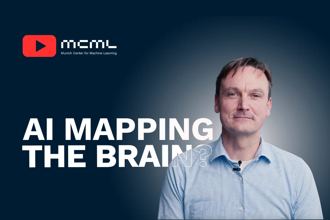
is Professor for AI in radiology at TU Munich.
He conducts research on novel AI algorithms for the analysis of medical images and their translation into clinical practice. He develops multimodal models for disease prediction and uses big data to train complex neural networks. Currently, he is focusing on the following challenges: (i) transparency of AI, (ii) integration of heterogeneous data, and (iii) generalization, bias, and fairness.
Team members @MCML
PostDocs
PhD Students

Jannik Lübberstedt
→ Group Christian Wachinger
Artificial Intelligence in Medical Imaging
Recent News @MCML
Publications @MCML
2026
[38]
F. Bongratz • Y. Li • S. Elbaroudy • C. Wachinger
Cortex-Grounded Diffusion Models for Brain Image Generation.
Preprint (Jan. 2026). arXiv
Cortex-Grounded Diffusion Models for Brain Image Generation.
Preprint (Jan. 2026). arXiv
[37]
M. Khateri • M. Ghahremani • S. Valencia • C. Jaimes • A. Sierra • J. Tohka • P. E. Grant • D. Karimi
USFetal: Tools for Fetal Brain Ultrasound Compounding.
Preprint (Jan. 2026). arXiv
USFetal: Tools for Fetal Brain Ultrasound Compounding.
Preprint (Jan. 2026). arXiv
2025
[36]

B. Schmitz-Koep • V. Schultz • F. Bongratz • A. Menegaux • M. Thalhammer • S. Schramm • S. H. Kim • C. Zimmer • C. Sorg • C. Wachinger • P. Giannakopoulos • M.-L. Montandon • C. Rodriguez • S. Haller • D. M. Hedderich
Longitudinal assessment of cortical thickness in healthy older individuals: a comparison between CAT12 and freesurfer.
Neuroradiology. Dec. 2025. DOI
Longitudinal assessment of cortical thickness in healthy older individuals: a comparison between CAT12 and freesurfer.
Neuroradiology. Dec. 2025. DOI
[35]
B. Jian • J. Pan • R. Jena • M. Ghahremani • H. B. Li • D. Rückert • C. Wachinger • B. Wiestler
Disentangling Progress in Medical Image Registration: Beyond Trend-Driven Architectures towards Domain-Specific Strategies.
Preprint (Dec. 2025). arXiv GitHub
Disentangling Progress in Medical Image Registration: Beyond Trend-Driven Architectures towards Domain-Specific Strategies.
Preprint (Dec. 2025). arXiv GitHub
[34]
N. Li • M. Ghahremani • B. Jian • P. T. Cervera • B. Wiestler • M. Makowski • C. Wachinger
Cross-Domain Semi-Supervised Organ Detection.
Preprint (Nov. 2025). URL
Cross-Domain Semi-Supervised Organ Detection.
Preprint (Nov. 2025). URL
[33]

T. N. Wolf • E. Kavak • F. Bongratz • C. Wachinger
SIC: Similarity-Based Interpretable Image Classification with Neural Networks.
ICCV 2025 - IEEE/CVF International Conference on Computer Vision. Honolulu, Hawai’i, Oct 19-23, 2025. To be published. Preprint available. URL GitHub
SIC: Similarity-Based Interpretable Image Classification with Neural Networks.
ICCV 2025 - IEEE/CVF International Conference on Computer Vision. Honolulu, Hawai’i, Oct 19-23, 2025. To be published. Preprint available. URL GitHub
[32]
E. O. Riedel • F. Bongratz • P. T. Zebhauser • M. Mühlau • C. Wachinger • D. M. Hedderich
Febrile Infection-Related Epilepsy Syndrome (FIRES) in a Young Adult: A Case Report Highlighting Advanced Neuroimaging.
Clinical Neuroradiology . Oct. 2025. DOI
Febrile Infection-Related Epilepsy Syndrome (FIRES) in a Young Adult: A Case Report Highlighting Advanced Neuroimaging.
Clinical Neuroradiology . Oct. 2025. DOI
[31]
Y. Li • R. Buchert • B. Schmitz-Koep • T. Grimmer • B. Ommer • D. M. Hedderich • I. Yakushev • C. Wachinger
Diffusion Bridge Networks Simulate Clinical-grade PET from MRI for Dementia Diagnostics.
Preprint (Oct. 2025). arXiv GitHub
Diffusion Bridge Networks Simulate Clinical-grade PET from MRI for Dementia Diagnostics.
Preprint (Oct. 2025). arXiv GitHub
[30]

F. Bongratz • T. N. Wolf • J. G. Ramon • C. Wachinger
X-SiT: Inherently Interpretable Surface Vision Transformers for Dementia Diagnosis.
MICCAI 2025 - 28th International Conference on Medical Image Computing and Computer Assisted Intervention. Daejeon, Republic of Korea, Sep 23-27, 2025. DOI
X-SiT: Inherently Interpretable Surface Vision Transformers for Dementia Diagnosis.
MICCAI 2025 - 28th International Conference on Medical Image Computing and Computer Assisted Intervention. Daejeon, Republic of Korea, Sep 23-27, 2025. DOI
[29]
P. Madlindl • F. Bongratz • C. Wachinger
Template-Based Cortical Surface Reconstruction with Minimal Energy Deformation.
ShapeMI @MICCAI 2025 - Workshop on Shape in Medical Imaging at the 28th International Conference on Medical Image Computing and Computer Assisted Intervention. Daejeon, Republic of Korea, Sep 23-27, 2025. DOI
Template-Based Cortical Surface Reconstruction with Minimal Energy Deformation.
ShapeMI @MICCAI 2025 - Workshop on Shape in Medical Imaging at the 28th International Conference on Medical Image Computing and Computer Assisted Intervention. Daejeon, Republic of Korea, Sep 23-27, 2025. DOI
[28]
I. Stoyanov • F. Bongratz • C. Wachinger
Spherical Brownian Bridge Diffusion Models for Conditional Cortical Thickness Forecasting.
ShapeMI @MICCAI 2025 - Workshop on Shape in Medical Imaging at the 28th International Conference on Medical Image Computing and Computer Assisted Intervention. Daejeon, Republic of Korea, Sep 23-27, 2025. DOI
Spherical Brownian Bridge Diffusion Models for Conditional Cortical Thickness Forecasting.
ShapeMI @MICCAI 2025 - Workshop on Shape in Medical Imaging at the 28th International Conference on Medical Image Computing and Computer Assisted Intervention. Daejeon, Republic of Korea, Sep 23-27, 2025. DOI
[27]

A. Taghipour • M. Ghahremani • M. Bennamoun • A. M. Rekavandi • H. Laga • F. Boussaid
Box It to Bind It: Unified Layout Control and Attribute Binding in Text-to-Image Diffusion Models.
IEEE Transactions on Multimedia Early Access. Sep. 2025. DOI
Box It to Bind It: Unified Layout Control and Attribute Binding in Text-to-Image Diffusion Models.
IEEE Transactions on Multimedia Early Access. Sep. 2025. DOI
[26]

C. Wachinger • D. M. Hedderich • M. Thalhammer • F. Bongratz
Individualized mapping of aberrant cortical thickness via stochastic cortical self-reconstruction.
Medical Image Analysis In press.Journal Pre-proof. Sep. 2025. DOI
Individualized mapping of aberrant cortical thickness via stochastic cortical self-reconstruction.
Medical Image Analysis In press.Journal Pre-proof. Sep. 2025. DOI
[25]
C. Liu • Y. Chen • H. Shi • J. Lu • B. Jian • J. Pan • L. Cai • J. Wang • Y. Zhang • J. Li • C. I. Bercea • C. Ouyang • C. Chen • Z. Xiong • B. Wiestler • C. Wachinger • D. Rückert • W. Bai • R. Arcucci
Does DINOv3 Set a New Medical Vision Standard?
Preprint (Sep. 2025). arXiv
Does DINOv3 Set a New Medical Vision Standard?
Preprint (Sep. 2025). arXiv
[24]

A. Taghipour • M. Ghahremani • M. Bennamoun • A. M. Rekavandi • Z. Li • H. Laga • F. Boussaid
Faster Image2Video Generation: A Closer Look at CLIP Image Embedding's Impact on Spatio-Temporal Cross-Attentions.
IEEE Access. Aug. 2025. DOI
Faster Image2Video Generation: A Closer Look at CLIP Image Embedding's Impact on Spatio-Temporal Cross-Attentions.
IEEE Access. Aug. 2025. DOI
[23]
J. Pan • B. Jian • P. Hager • Y. Zhang • C. Liu • F. Jungmann • H. B. Li • C. You • J. Wu • J. Zhu • F. Liu • Y. Liu • N. Bubeck • C. Wachinger • C. Chen • Z. Gong • C. Ouyang • G. Kaissis • B. Wiestler • D. Rückert
Beyond Benchmarks: Dynamic, Automatic And Systematic Red-Teaming Agents For Trustworthy Medical Language Models.
Preprint (Aug. 2025). arXiv
Beyond Benchmarks: Dynamic, Automatic And Systematic Red-Teaming Agents For Trustworthy Medical Language Models.
Preprint (Aug. 2025). arXiv
[22]
Y. Li • M. Ghahremani • C. Wachinger
MedBridge: Bridging Foundation Vision-Language Models to Medical Image Diagnosis in Chest X-Ray.
ICVSS 2025 - International Computer Vision Summer School: Computer Vision for Spatial Intelligence. Sicily, Italy, Jul 06-12, 2025. To be published. Preprint available. arXiv GitHub
MedBridge: Bridging Foundation Vision-Language Models to Medical Image Diagnosis in Chest X-Ray.
ICVSS 2025 - International Computer Vision Summer School: Computer Vision for Spatial Intelligence. Sicily, Italy, Jul 06-12, 2025. To be published. Preprint available. arXiv GitHub
[21]
E. Kavak • T. N. Wolf • C. Wachinger
DISCO: Mitigating Bias in Deep Learning with Conditional Distance Correlation.
Preprint (Jun. 2025). arXiv
DISCO: Mitigating Bias in Deep Learning with Conditional Distance Correlation.
Preprint (Jun. 2025). arXiv
[20]
F. Bongratz • Y. Li • S. Elbaroudy • C. Wachinger
3D Shape-to-Image Brownian Bridge Diffusion for Brain MRI Synthesis from Cortical Surfaces.
IPMI 2025 - Information Processing in Medical Imaging. Kos Island, Greece, May 25-30, 2025. DOI GitHub
3D Shape-to-Image Brownian Bridge Diffusion for Brain MRI Synthesis from Cortical Surfaces.
IPMI 2025 - Information Processing in Medical Imaging. Kos Island, Greece, May 25-30, 2025. DOI GitHub
[19]

A.-M. Rickmann • F. Bongratz • C. Wachinger
Vertex Correspondence and Self-Intersection Reduction in Cortical Surface Reconstruction.
IEEE Transactions on Medical Imaging Early Access. Apr. 2025. DOI
Vertex Correspondence and Self-Intersection Reduction in Cortical Surface Reconstruction.
IEEE Transactions on Medical Imaging Early Access. Apr. 2025. DOI
[18]

Y. Li • M. Ghahremani • Y. Wally • C. Wachinger
DiaMond: Dementia Diagnosis with Multi-Modal Vision Transformers Using MRI and PET.
WACV 2025 - IEEE/CVF Winter Conference on Applications of Computer Vision. Tucson, AZ, USA, Feb 28-Mar 04, 2025. DOI
DiaMond: Dementia Diagnosis with Multi-Modal Vision Transformers Using MRI and PET.
WACV 2025 - IEEE/CVF Winter Conference on Applications of Computer Vision. Tucson, AZ, USA, Feb 28-Mar 04, 2025. DOI
[17]

M. Ghahremani • B. R. Ernhofer • J. Wang • M. Makowski • C. Wachinger
Organ-DETR: Organ Detection via Transformers.
IEEE Transactions on Medical Imaging Early Access. Feb. 2025. DOI URL
Organ-DETR: Organ Detection via Transformers.
IEEE Transactions on Medical Imaging Early Access. Feb. 2025. DOI URL
[16]
F. Bongratz • J. Fecht • A.-M. Rickmann • C. Wachinger
V2C-Long: Longitudinal Cortex Reconstruction with Spatiotemporal Correspondence.
Preprint (Feb. 2025). arXiv
V2C-Long: Longitudinal Cortex Reconstruction with Spatiotemporal Correspondence.
Preprint (Feb. 2025). arXiv
[15]
B. Jian • J. Pan • Y. Li • F. Bongratz • R. Li • D. Rückert • B. Wiestler • C. Wachinger
TimeFlow: Longitudinal Brain Image Registration and Aging Progression Analysis.
Preprint (Jan. 2025). arXiv
TimeFlow: Longitudinal Brain Image Registration and Aging Progression Analysis.
Preprint (Jan. 2025). arXiv
2024
[14]

J. Wang • M. Ghahremani • Y. Li • B. Ommer • C. Wachinger
Stable-Pose: Leveraging Transformers for Pose-Guided Text-to-Image Generation.
NeurIPS 2024 - 38th Conference on Neural Information Processing Systems. Vancouver, Canada, Dec 10-15, 2024. URL GitHub
Stable-Pose: Leveraging Transformers for Pose-Guided Text-to-Image Generation.
NeurIPS 2024 - 38th Conference on Neural Information Processing Systems. Vancouver, Canada, Dec 10-15, 2024. URL GitHub
[13]
F. Bongratz • M. Karmann • A. Holz • M. Bonhoeffer • V. Neumaier • S. Deli • B. Schmitz-Koep • C. Zimmer • C. Sorg • M. Thalhammer • D. M. Hedderich • C. Wachinger
MLV2-Net: Rater-Based Majority-Label Voting for Consistent Meningeal Lymphatic Vessel Segmentation.
Preprint (Nov. 2024). arXiv
MLV2-Net: Rater-Based Majority-Label Voting for Consistent Meningeal Lymphatic Vessel Segmentation.
Preprint (Nov. 2024). arXiv
[12]

Y. Li • I. Yakushev • D. M. Hedderich • C. Wachinger
PASTA: Pathology-Aware MRI to PET Cross-Modal Translation with Diffusion Models.
MICCAI 2024 - 27th International Conference on Medical Image Computing and Computer Assisted Intervention. Marrakesh, Morocco, Oct 06-10, 2024. DOI GitHub
PASTA: Pathology-Aware MRI to PET Cross-Modal Translation with Diffusion Models.
MICCAI 2024 - 27th International Conference on Medical Image Computing and Computer Assisted Intervention. Marrakesh, Morocco, Oct 06-10, 2024. DOI GitHub
[11]
B. Jian • J. Pan • M. Ghahremani • D. Rückert • C. Wachinger • B. Wiestler
Mamba? Catch The Hype Or Rethink What Really Helps for Image Registration.
WBIR @MICCAI 2024 - 11th International Workshop on Biomedical Image Registration at the 27th International Conference on Medical Image Computing and Computer Assisted Intervention. Marrakesh, Morocco, Oct 06-10, 2024. DOI
Mamba? Catch The Hype Or Rethink What Really Helps for Image Registration.
WBIR @MICCAI 2024 - 11th International Workshop on Biomedical Image Registration at the 27th International Conference on Medical Image Computing and Computer Assisted Intervention. Marrakesh, Morocco, Oct 06-10, 2024. DOI
[10]
Y. Li • T. N. Wolf • S. Pölsterl • I. Yakushev • D. M. Hedderich • C. Wachinger
From Barlow Twins to Triplet Training: Differentiating Dementia with Limited Data.
MIDL 2024 - Medical Imaging with Deep Learning. Paris, France, Jul 03-05, 2024. URL
From Barlow Twins to Triplet Training: Differentiating Dementia with Limited Data.
MIDL 2024 - Medical Imaging with Deep Learning. Paris, France, Jul 03-05, 2024. URL
[9]
F. Bongratz • V. Golkov • L. Mautner • L. Della Libera • F. Heetmeyer • F. Czaja • J. Rodemann • D. Cremers
How to Choose a Reinforcement-Learning Algorithm.
Preprint (Jul. 2024). arXiv GitHub
How to Choose a Reinforcement-Learning Algorithm.
Preprint (Jul. 2024). arXiv GitHub
[8]

M. Ghahremani • M. Khateri • B. Jian • B. Wiestler • E. Adeli • C. Wachinger
H-ViT: A Hierarchical Vision Transformer for Deformable Image Registration.
CVPR 2024 - IEEE/CVF Conference on Computer Vision and Pattern Recognition. Seattle, WA, USA, Jun 17-21, 2024. DOI
H-ViT: A Hierarchical Vision Transformer for Deformable Image Registration.
CVPR 2024 - IEEE/CVF Conference on Computer Vision and Pattern Recognition. Seattle, WA, USA, Jun 17-21, 2024. DOI
[7]
C. Wachinger • D. M. Hedderich • F. Bongratz
Stochastic Cortical Self-Reconstruction.
Preprint (Mar. 2024). arXiv
Stochastic Cortical Self-Reconstruction.
Preprint (Mar. 2024). arXiv
[6]

T. N. Wolf • F. Bongratz • A.-M. Rickmann • S. Pölsterl • C. Wachinger
Keep the Faith: Faithful Explanations in Convolutional Neural Networks for Case-Based Reasoning.
AAAI 2024 - 38th Conference on Artificial Intelligence. Vancouver, Canada, Feb 20-27, 2024. DOI
Keep the Faith: Faithful Explanations in Convolutional Neural Networks for Case-Based Reasoning.
AAAI 2024 - 38th Conference on Artificial Intelligence. Vancouver, Canada, Feb 20-27, 2024. DOI
[5]

F. Bongratz • A.-M. Rickmann • C. Wachinger
Neural deformation fields for template-based reconstruction of cortical surfaces from MRI.
Medical Image Analysis 93. Jan. 2024. DOI
Neural deformation fields for template-based reconstruction of cortical surfaces from MRI.
Medical Image Analysis 93. Jan. 2024. DOI
2023
[4]

M. Ghahremani • C. Wachinger
RegBN: Batch Normalization of Multimodal Data with Regularization.
NeurIPS 2023 - 37th Conference on Neural Information Processing Systems. New Orleans, LA, USA, Dec 10-16, 2023. URL GitHub
RegBN: Batch Normalization of Multimodal Data with Regularization.
NeurIPS 2023 - 37th Conference on Neural Information Processing Systems. New Orleans, LA, USA, Dec 10-16, 2023. URL GitHub
[3]

C. Wachinger • T. N. Wolf • S. Pölsterl
Deep learning for the prediction of type 2 diabetes mellitus from neck-to-knee Dixon MRI in the UK biobank.
Heliyon 9.11. Nov. 2023. DOI
Deep learning for the prediction of type 2 diabetes mellitus from neck-to-knee Dixon MRI in the UK biobank.
Heliyon 9.11. Nov. 2023. DOI
[2]

F. Bongratz • A.-M. Rickmann • C. Wachinger
Abdominal organ segmentation via deep diffeomorphic mesh deformations.
Scientific Reports 13.1. Oct. 2023. DOI
Abdominal organ segmentation via deep diffeomorphic mesh deformations.
Scientific Reports 13.1. Oct. 2023. DOI
2021
[1]
P. Kopper • S. Pölsterl • C. Wachinger • B. Bischl • A. Bender • D. Rügamer
Semi-Structured Deep Piecewise Exponential Models.
AAAI-SPACA 2021 - AAAI Spring Symposium Series on Survival Prediction: Algorithms, Challenges and Applications. Palo Alto, California, USA, Mar 21-24, 2021. PDF
Semi-Structured Deep Piecewise Exponential Models.
AAAI-SPACA 2021 - AAAI Spring Symposium Series on Survival Prediction: Algorithms, Challenges and Applications. Palo Alto, California, USA, Mar 21-24, 2021. PDF
©all images: LMU | TUM

