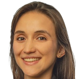
Research Group Martin Menten
Martin Menten
leads the MCML Junior Research Group ‘AI for Vision’ at TU Munich.
He and his research group specialize in machine learning for medical imaging. Their research focuses on weakly and self-supervised learning to address data scarcity in healthcare and the integration of multimodal clinical data with medical images. In particular, they are interested in the development and application of machine learning and computer vision algorithms in the field of ophthalmology. Funded by the DFG, the group investigates new research directions that complement and extend MCML’s focus while remaining closely connected to the center.
Team members @MCML
PhD Students
Recent News @MCML
Publications @MCML
2025
[21]
L. Lux • A. H. Berger • M. R. Tricas • R. Rosen • A. E. Fayed • S. Sivaprasada • L. Kreitner • J. Weidner • M. J. Menten • D. Rückert • J. C. Paetzold
Interpretable Retinal Disease Prediction Using Biology-Informed Heterogeneous Graph Representations.
Preprint (Nov. 2025). arXiv
Interpretable Retinal Disease Prediction Using Biology-Informed Heterogeneous Graph Representations.
Preprint (Nov. 2025). arXiv
[20]

Y. Bi • L. Huang • R. Clarenbach • R. Ghotbi • A. Karlas • N. Navab • Z. Jiang
Synomaly noise and multi-stage diffusion: A novel approach for unsupervised anomaly detection in medical images.
Medical Image Analysis 105.103737. Oct. 2025. DOI GitHub
Synomaly noise and multi-stage diffusion: A novel approach for unsupervised anomaly detection in medical images.
Medical Image Analysis 105.103737. Oct. 2025. DOI GitHub
[19]
L. Kreitner • P. Hager • J. Mengedoht • G. Kaissis • D. Rückert • M. J. Menten
Efficient numeracy in language models through single-token number embeddings.
Preprint (Oct. 2025). arXiv
Efficient numeracy in language models through single-token number embeddings.
Preprint (Oct. 2025). arXiv
[18]
M. Hartenberger • H. Ayaz • F. Ozlugedik • C. Caredda • L. Giannoni • F. Lange • L. Lux • J. Weidner • A. Berger • F. Kofler • M. J. Menten • B. Montcel • I. Tachtsidis • D. Rückert • I. Ezhov
Redefining spectral unmixing for in-vivo brain tissue analysis from hyperspectral imaging.
CMMCA @MICCAI 2025 - Workshop on Computational Mathematics Modeling in Cancer Analysis at 28th International Conference on Medical Image Computing and Computer Assisted Intervention. Daejeon, Republic of Korea, Sep 23-27, 2025. DOI
[17]

S. Starck • V. Sideri-Lampretsa • B. Kainz • M. J. Menten • T. T. Mueller • D. Rückert
Diff-Def: Diffusion-Generated Deformation Fields for Conditional Atlases.
IEEE Transactions on Medical Imaging Early Access. Aug. 2025. DOI
Diff-Def: Diffusion-Generated Deformation Fields for Conditional Atlases.
IEEE Transactions on Medical Imaging Early Access. Aug. 2025. DOI
[16]

R. Holland • T. R. P. Taylor • C. Holmes • S. Riedl • J. Mai • M. Patsiamanidi • D. Mitsopoulou • P. Hager • P. Müller • J. C. Paetzold • H. P. N. Scholl • H. Bogunović • U. Schmidt-Erfurth • D. Rückert • S. Sivaprasad • A. J. Lotery • M. J. Menten • O. b. o. t. PINNACLE consortium
Specialized curricula for training vision language models in retinal image analysis.
npj Digital Medicine 8.532. Aug. 2025. DOI
Specialized curricula for training vision language models in retinal image analysis.
npj Digital Medicine 8.532. Aug. 2025. DOI
[15]
K. Yang • F. Musio • Y. Ma • N. Juchler • J. C. Paetzold • R. Al-Maskari • L. Höher • H. B. Li • I. E. Hamamci • A. Sekuboyina • S. Shit • H. Huang • C. Prabhakar • E. de la Rosa • B. Wittmann • D. Waldmannstetter • F. Kofler • F. Navarro • M. J. Menten • I. Ezhov • D. Rückert • I. N. Vos • Y. M. Ruigrok • B. K. Velthuis • H. J. Kuijf • P. Shi • W. Liu • T. Ma • M. R. Rokuss • Y. Kirchhoff • F. Isensee • K. Maier-Hein • C. Zhu • H. Zhao • P. Bijlenga • J. Hämmerli • C. Wurster • L. Westphal • J. Bisschop • E. Colombo • H. Baazaoui • H.-L. Handelsmann • A. Makmur • J. Hallinan • A. Soundararajan • B. Wiestler • J. S. Kirschke • R. Wiest • E. Montagnon • L. Letourneau-Guillon • K. Oh • D. Lee • A. Hilbert • O. U. Aydin • D. Rallios • J. Rieger • S. Tanioka • A. Koch • D. Frey • A. Qayyum • M. Mazher • S. Niederer • N. Disch • J. Holzschuh • D. LaBella • F. Galati • D. Falcetta • M. A. Zuluaga • C. Lin • H. Zhao • Z. Zhang • M. Zhang • X. You • H. Zhang • G.-Z. Yang • Y. Gu • S. Ra • J. Hwang • H. Park • J. Chen • M. Wodzinski • H. Müller • N. Mansouri • F. Autrusseau • C. Yalçin • R. E. Hamadache • C. Lisazo • J. Salvi • A. Casamitjana • X. Lladó • U. M. Lal-Trehan Estrada • V. Abramova • L. Giancardo • A. Oliver • P. Casademunt • A. Galdran • M. Delucchi • J. Liu • H. Huang • Y. Cui • Z. Lin • Y. Liu • S. Zhu • T. R. Patel • A. H. Siddiqui • V. M. Tutino • M. Orouskhani • H. Wang • M. Mossa-Basha • Y. Sato • S. Hirsch • S. Wegener • B. Menze
Benchmarking the CoW with the TopCoW Challenge: Topology-Aware Anatomical Segmentation of the Circle of Willis for CTA and MRA.
Preprint (Jul. 2025). arXiv
Benchmarking the CoW with the TopCoW Challenge: Topology-Aware Anatomical Segmentation of the Circle of Willis for CTA and MRA.
Preprint (Jul. 2025). arXiv
[14]

D. Mildenberger • P. Hager • D. Rückert • M. J. Menten
A Tale of Two Classes: Adapting Supervised Contrastive Learning to Binary Imbalanced Datasets.
CVPR 2025 - IEEE/CVF Conference on Computer Vision and Pattern Recognition. Nashville, TN, USA, Jun 11-15, 2025. DOI
A Tale of Two Classes: Adapting Supervised Contrastive Learning to Binary Imbalanced Datasets.
CVPR 2025 - IEEE/CVF Conference on Computer Vision and Pattern Recognition. Nashville, TN, USA, Jun 11-15, 2025. DOI
[13]
A. H. Berger • L. Lux • A. Weers • M. J. Menten • D. Rückert • J. C. Paetzold
Pitfalls of topology-aware image segmentation.
IPMI 2025 - Information Processing in Medical Imaging. Kos Island, Greece, May 25-30, 2025. DOI
Pitfalls of topology-aware image segmentation.
IPMI 2025 - Information Processing in Medical Imaging. Kos Island, Greece, May 25-30, 2025. DOI
[12]
L. D. Reyes Vargas • M. J. Menten • J. C. Paetzold • N. Navab • M. F. Azampour
Skelite: Compact Neural Networks for Efficient Iterative Skeletonization.
IPMI 2025 - Information Processing in Medical Imaging. Kos Island, Greece, May 25-30, 2025. DOI
Skelite: Compact Neural Networks for Efficient Iterative Skeletonization.
IPMI 2025 - Information Processing in Medical Imaging. Kos Island, Greece, May 25-30, 2025. DOI
[11]
S. Lockfisch • K. Schwethelm • M. J. Menten • R. Braren • D. Rückert • A. Ziller • G. Kaissis
On Arbitrary Predictions from Equally Valid Models.
Preprint (May. 2025). arXiv
On Arbitrary Predictions from Equally Valid Models.
Preprint (May. 2025). arXiv
[10]

L. Lux • A. H. Berger • A. Weers • N. Stucki • D. Rückert • U. Bauer • J. C. Paetzold
Topograph: An efficient Graph-Based Framework for Strictly Topology Preserving Image Segmentation.
ICLR 2025 - 13th International Conference on Learning Representations. Singapore, Apr 24-28, 2025. URL
Topograph: An efficient Graph-Based Framework for Strictly Topology Preserving Image Segmentation.
ICLR 2025 - 13th International Conference on Learning Representations. Singapore, Apr 24-28, 2025. URL
[9]

Ö. Turgut • P. Müller • P. Hager • S. Shit • S. Starck • M. J. Menten • E. Martens • D. Rückert
Unlocking the diagnostic potential of electrocardiograms through information transfer from cardiac magnetic resonance imaging.
Medical Image Analysis 101.103451. Apr. 2025. DOI GitHub
Unlocking the diagnostic potential of electrocardiograms through information transfer from cardiac magnetic resonance imaging.
Medical Image Analysis 101.103451. Apr. 2025. DOI GitHub
[8]
A. Weers • A. H. Berger • L. Lux • P. J. Schüffler • D. Rückert • J. C. Paetzold
From Pixels to Histopathology: A Graph-Based Framework for Interpretable Whole Slide Image Analysis.
Preprint (Mar. 2025). arXiv GitHub
From Pixels to Histopathology: A Graph-Based Framework for Interpretable Whole Slide Image Analysis.
Preprint (Mar. 2025). arXiv GitHub
[7]

A. H. Berger • L. Lux • S. Shit • I. Ezhov • G. Kaissis • M. J. Menten • D. Rückert • J. C. Paetzold
Cross-Domain and Cross-Dimension Learning for Image-to-Graph Transformers.
WACV 2025 - IEEE/CVF Winter Conference on Applications of Computer Vision. Tucson, AZ, USA, Feb 28-Mar 04, 2025. DOI
Cross-Domain and Cross-Dimension Learning for Image-to-Graph Transformers.
WACV 2025 - IEEE/CVF Winter Conference on Applications of Computer Vision. Tucson, AZ, USA, Feb 28-Mar 04, 2025. DOI
2024
[6]
L. Lux • A. H. Berger • M. Romeo-Tricas • M. J. Menten • D. Rückert • J. C. Paetzold
Exploring Graphs as Data Representation for Disease Classification in Ophthalmology.
GRAIL @MICCAI 2024 - 6th Workshop on GRaphs in biomedicAl Image anaLysis at the 27th International Conference on Medical Image Computing and Computer Assisted Intervention. Marrakesh, Morocco, Oct 06-10, 2024. DOI URL
[5]

R. Wicklein • L. Kreitner • A. Wild • L. Aly • D. Rückert • B. Hemmer • T. Korn • M. J. Menten • B. Knier
Retinal small vessel pathology is associated with disease burden in multiple sclerosis.
Multiple Sclerosis Journal 30.7. Jun. 2024. DOI
Retinal small vessel pathology is associated with disease burden in multiple sclerosis.
Multiple Sclerosis Journal 30.7. Jun. 2024. DOI
[4]

L. Kreitner • J. C. Paetzold • N. Rauch • C. Chen • A. M. Hagag • A. E. Fayed • S. Sivaprasad • S. Rausch • J. Weichsel • B. H. Menze • M. Harders • B. Knier • D. Rückert • M. J. Menten
Synthetic Optical Coherence Tomography Angiographs for Detailed Retinal Vessel Segmentation Without Human Annotations.
IEEE Transactions on Medical Imaging 43.6. Jan. 2024. DOI
Synthetic Optical Coherence Tomography Angiographs for Detailed Retinal Vessel Segmentation Without Human Annotations.
IEEE Transactions on Medical Imaging 43.6. Jan. 2024. DOI
2023
[3]
D. Scholz • B. Wiestler • D. Rückert • M. J. Menten
Metrics to Quantify Global Consistency in Synthetic Medical Images.
DGM4 @MICCAI 2023 - 3rd International Workshop on Deep Generative Models at the 26th International Conference on Medical Image Computing and Computer Assisted Intervention. Vancouver, Canada, Oct 08-12, 2023. DOI
Metrics to Quantify Global Consistency in Synthetic Medical Images.
DGM4 @MICCAI 2023 - 3rd International Workshop on Deep Generative Models at the 26th International Conference on Medical Image Computing and Computer Assisted Intervention. Vancouver, Canada, Oct 08-12, 2023. DOI
[2]

R. Holland • O. Leingang • C. Holmes • P. Anders • R. Kaye • S. Riedl • J. C. Paetzold • I. Ezhov • H. Bogunović • U. Schmidt-Erfurth • H. P. N. Scholl • S. Sivaprasad • A. J. Lotery • D. Rückert • M. J. Menten
Clustering Disease Trajectories in Contrastive Feature Space for Biomarker Proposal in Age-Related Macular Degeneration.
MICCAI 2023 - 26th International Conference on Medical Image Computing and Computer Assisted Intervention. Vancouver, Canada, Oct 08-12, 2023. DOI
Clustering Disease Trajectories in Contrastive Feature Space for Biomarker Proposal in Age-Related Macular Degeneration.
MICCAI 2023 - 26th International Conference on Medical Image Computing and Computer Assisted Intervention. Vancouver, Canada, Oct 08-12, 2023. DOI
[1]

M. J. Menten • J. C. Paetzold • V. A. Zimmer • S. Shit • I. Ezhov • R. Holland • M. Probst • J. A. Schnabel • D. Rückert
A Skeletonization Algorithm for Gradient-Based Optimization.
ICCV 2023 - IEEE/CVF International Conference on Computer Vision. Paris, France, Oct 02-06, 2023. DOI
A Skeletonization Algorithm for Gradient-Based Optimization.
ICCV 2023 - IEEE/CVF International Conference on Computer Vision. Paris, France, Oct 02-06, 2023. DOI
©all images: LMU | TUM

