Research Group Michael Ingrisch

Michael Ingrisch
Prof. Dr.
Core PI
Michael Ingrisch
leads the group for Clinical Data Science at the Department of Radiology at LMU Munich.
His team employs advanced statistics, machine learning and computer vision techniques in the context of clinical radiology to enable fast and precise AI-supported diagnosis and prognostication. The research areas focus on applying computer vision techniques in radiology for diagnosis and prognosis, as well as using biostatistical methods to rigorously analyze clinical data. Additionally, the work includes leveraging large language models for clinical text analysis and developing multimodal deep learning models that integrate diverse data types, such as imaging and text, to improve AI model accuracy and applicability.
Team members @MCML
PostDocs

Boj Friedrich Hoppe
Dr.
→ Group Michael Ingrisch
Clinical Data Science in Radiology
Katharina Jeblick
Dr.
→ Group Michael Ingrisch
Clinical Data Science in Radiology

Balthasar Schachtner
Dr.
→ Group Michael Ingrisch
Clinical Data Science in Radiology
PhD Students

Jakob Dexl
→ Group Michael Ingrisch
Clinical Data Science in Radiology

Theresa Stüber
→ Group Michael Ingrisch
Clinical Data Science in Radiology
Johanna Topalis
→ Group Michael Ingrisch
Clinical Data Science in Radiology
Recent News @MCML

Publications @MCML
2026
Prognostic and predictive value of radiomics-based imaging features in patients with colorectal liver metastasis receiving radioembolisation in first-line setting.
European Journal of Radiology Open 16. Jun. 2026. DOI
The autoPET3 Challenge: Automated Lesion Segmentation in Whole-Body PET/CT – Multitracer Multicenter Generalization.
Preprint (May. 2026). arXiv GitHub
Artificial intelligence for TNM staging in NSCLC: a critical appraisal of segmentation utility in [1⁸F]FDG PET/CT.
European Journal of Nuclear Medicine and Molecular Imaging 53. Apr. 2026. DOI
The architectural gap in clinical artificial intelligence.
The Lancet Digital Healths 8.2. Apr. 2026. DOI
The effect of medical explanations from large language models on diagnostic accuracy in radiology.
npj Digital Medicine 9.333. Apr. 2026. DOI
Fully automated segmentation of [18F]FDG- and PSMA-PET/CT images via data-centric Deep-Learning.
Preprint (Apr. 2026). DOI GitHub
AutoPET Challenge on Fully Automated Lesion Segmentation in Oncologic PET/CT Imaging, Part 2: Domain Generalization.
Journal of Nuclear Medicine 67.3. Mar. 2026. DOI
Reporting checklist for foundation and large language models in medical research (REFINE): an international consensus guideline.
Diagnostic and Interventional Radiology. Feb. 2026. DOI
2025
Fast machine learning image reconstruction of radially undersampled k-space data for low-latency real-time MRI.
PLOS One 20.11. Nov. 2025. DOI
3D deep learning-based muscle volume quantification from thoracic CT as a surrogate for DXA-Derived appendicular muscle mass in older adults.
Aging Clinical and Experimental Research 37.296. Oct. 2025. DOI
Multi-Task Deep Learning for Head and Neck Cancer: Segmentation, Survival Prediction, and HPV Classification in the HECKTOR 2025 Challenge.
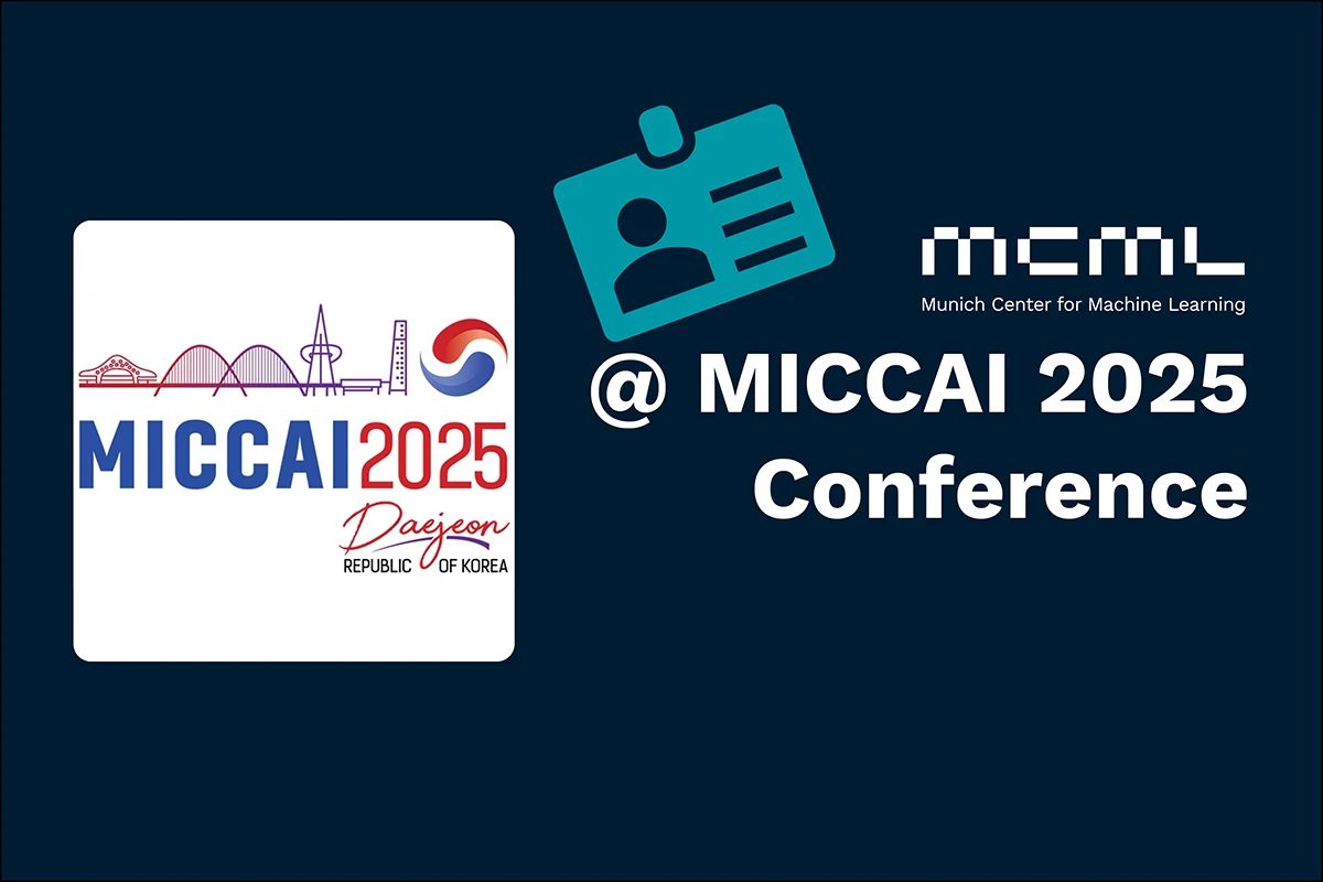
HECKTOR @MICCAI 2025 - 4th Head and Neck Cancer Tumor Lesion Segmentation, Diagnosis and Prognosis Challenge at the 28th International Conference on Medical Image Computing and Computer Assisted Intervention. Daejeon, Republic of Korea, Sep 23-27, 2025. URL GitHub
Preventing Sensitive Information Leakage via Post-hoc Orthogonalization with Application to Chest Radiograph Embeddings.
PAKDD 2025 - 29th Pacific-Asia Conference on Knowledge Discovery and Data Mining. Sydney, Australia, Jun 10-13, 2025. DOI GitHub
PARADIM: A Platform to Support Research at the Interface of Data Science and Medical Imaging.
Journal of Imaging Informatics in Medicine. Jun. 2025. DOI
Radiomics-based differentiation of upper urinary tract urothelial and renal cell carcinoma in preoperative computed tomography datasets.
BMC Medical Imaging 25.196. May. 2025. DOI
Optimizing lower extremity CT angiography: A prospective study of individualized vs. fixed post-trigger delays in bolus tracking.
European Journal of Radiology 185.112009. Apr. 2025. DOI
Breast cancer risk prediction using background parenchymal enhancement, radiomics, and symmetry features on MRI.
SPIE 2025 - SPIE Medical Imaging: Computer-Aided Diagnosis. San Diego, CA, USA, Feb 16-21, 2025. DOI
Pitfalls with anomaly detection for breast cancer risk prediction.
SPIE 2025 - SPIE Medical Imaging: Computer-Aided Diagnosis. San Diego, CA, USA, Feb 16-21, 2025. DOI
Replication study of PD-L1 status prediction in NSCLC using PET/CT radiomics.
European Journal of Radiology 183.111825. Feb. 2025. DOI
Effect of artificial intelligence-aided differentiation of adenomatous and non-adenomatous colorectal polyps at CT colonography on radiologists’ therapy management.
European Radiology 35.7. Jan. 2025. DOI
A probability model for estimating age in young individuals relative to key legal thresholds: 15, 18 or 21-year.
International Journal of Legal Medicine 139.1. Jan. 2025. DOI
Post-Training Network Compression for 3D Medical Image Segmentation: Reducing Computational Efforts via Tucker Decomposition.
Radiology: Artificial Intelligence 7.2. Jan. 2025. DOI
2024
Artificial intelligence–based rapid brain volumetry substantially improves differential diagnosis in dementia.
Alzheimer’s and Dementia 16.e70037. Oct. 2024. DOI
Software-assisted structured reporting and semi-automated TNM classification for NSCLC staging in a multicenter proof of concept study.
Insights into Imaging 15.258. Oct. 2024. DOI
Results from the autoPET challenge on fully automated lesion segmentation in oncologic PET/CT imaging.
Nature Machine Intelligence 6. Oct. 2024. DOI
Automatische ICD-10-Codierung.
Die Radiologie 64. Aug. 2024. DOI
MRI-based ventilation and perfusion imaging to predict radiation-induced pneumonitis in lung tumor patients at a 0.35T MR-Linac.
Radiotherapy and Oncology. Aug. 2024. DOI
Towards Aleatoric and Epistemic Uncertainty in Medical Image Classification.
AIME 2024 - 22nd International Conference on Artificial Intelligence in Medicine. Salt Lake City, UT, USA, Jul 09-12, 2024. DOI
Using Ventilation and Perfusion MRI at a 0.35 T MR-Linac to Predict Radiation-Induced Pneumonitis in Lung Cancer Patients.
ISMRM 2024 - International Society for Magnetic Resonance in Medicine Annual Meeting. Singapore, May 04-09, 2024. URL
ChatGPT makes medicine easy to swallow: an exploratory case study on simplified radiology reports.
European Radiology 34. May. 2024. DOI
Application of machine learning in CT colonography and radiological age assessment: enhancing traditional diagnostics in radiology.
Dissertation LMU München. Mar. 2024. DOI
Computer vision-based guidance tool for correct radiographic hand positioning.
SPIE 2024 - SPIE Medical Imaging: Image Perception, Observer Performance, and Technology Assessment. San Diego, CA, USA, Feb 18-22, 2024. DOI
Deformable current-prior registration of DCE breast MR images on multi-site data.
SPIE 2024 - SPIE Medical Imaging: Image Processing. San Diego, CA, USA, Feb 18-22, 2024. DOI
Constrained Probabilistic Mask Learning for Task-specific Undersampled MRI Reconstruction.
WACV 2024 - IEEE/CVF Winter Conference on Applications of Computer Vision. Waikoloa, Hawaii, Jan 04-08, 2024. DOI
Radiological age assessment based on clavicle ossification in CT: enhanced accuracy through deep learning.
International Journal of Legal Medicine. Jan. 2024. DOI
2023
A comprehensive machine learning benchmark study for radiomics-based survival analysis of CT imaging data in patients with hepatic metastases of CRC.
Investigative Radiology 58.12. Dec. 2023. DOI
Post-hoc Orthogonalization for Mitigation of Protected Feature Bias in CXR Embeddings.
Preprint (Nov. 2023). arXiv
Revitalize the Potential of Radiomics: Interpretation and Feature Stability in Medical Imaging Analyses through Groupwise Feature Importance.
LB-D-DC 2023 @xAI 2023 - Late-breaking Work, Demos and Doctoral Consortium at the 1st World Conference on eXplainable Artificial Intelligence. Lisbon, Portugal, Jul 26-28, 2023. PDF
Cascaded Latent Diffusion Models for High-Resolution Chest X-ray Synthesis.
PAKDD 2023 - 27th Pacific-Asia Conference on Knowledge Discovery and Data Mining. Osaka, Japan, May 25-28, 2023. DOI
Robust evaluation of contrast-enhanced imaging for perfusion quantification.
Dissertation LMU München. May. 2023. DOI
2022
Implicit Embeddings via GAN Inversion for High Resolution Chest Radiographs.
MAD @MICCAI 2022 - 1st Workshop on Medical Applications with Disentanglements at the 25th International Conference on Medical Image Computing and Computer Assisted Intervention. Singapore, Sep 18-22, 2022. DOI
2021
Survival-oriented embeddings for improving accessibility to complex data structures.
Bridging the Gap: from ML Research to Clinical Practice @NeurIPS 2021 - Workshop on Bridging the Gap: from Machine Learning Research to Clinical Practice at the 35th Conference on Neural Information Processing Systems. Virtual, Dec 06-14, 2021. arXiv
Towards modelling hazard factors in unstructured data spaces using gradient-based latent interpolation.
Deep Generative Models and Downstream Applications @NeurIPS 2021 - Workshop on Deep Generative Models and Downstream Applications at the 35th Conference on Neural Information Processing Systems. Virtual, Dec 06-14, 2021. PDF
Bi-Centric Independent Validation of Outcome Prediction after Radioembolization of Primary and Secondary Liver Cancer.
Journal of Clinical Medicine 10.16. Aug. 2021. DOI
©all images: LMU | TUM