Research Group Peter Schüffler
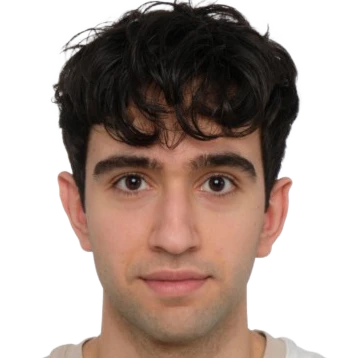
Peter Schüffler
is Professor for Computational Pathology at TU Munich.
His field of research is the area of digital and computational pathology. This includes novel machine learning approaches for the detection, segmentation and grading of cancer in pathology images, prediction of prognostic markers and outcome prediction (e.g. treatment response). Further, he investigates the efficient visualization of high-resolution digital pathology images, automated QA, new ergonomics for pathologists, and holistic integration of digital systems for clinics, research and education.
Team members @MCML
PostDocs
PhD Students
Recent News @MCML
Publications @MCML
2026
[20]

M. Fischer • A. Muckenhuber • R. Peretzke • L. Farah • C. Ulrich • S. Ziegler • P. Schader • L. Feineis • H. Gao • S. Xiao • M. Götz • M. Nolden • K. Steiger • J. T. Sieveke • L. Endrös • R. Braren • J. Kleesiek • P. J. Schüffler • P. Neher • K. Maier-Hein
Contrastive virtual staining enhances deep learning-based PDAC subtyping from H&E-stained tissue cores.
Journal of Pathology 268.1. Jan. 2026. DOI
Contrastive virtual staining enhances deep learning-based PDAC subtyping from H&E-stained tissue cores.
Journal of Pathology 268.1. Jan. 2026. DOI
2025
[19]
R. Fani • R. Al Attrach • D. Restrepo • Y. Jia • L. A. Celi • P. J. Schüffler
Coefficient of Variation Masking: A Volatility-Aware Strategy for EHR Foundation Models.
ML4H 2025 - Machine Learning for Health Symposium. San Diego, CA, USA, Dec 01-02, 2025. To be published. Preprint available. arXiv
Coefficient of Variation Masking: A Volatility-Aware Strategy for EHR Foundation Models.
ML4H 2025 - Machine Learning for Health Symposium. San Diego, CA, USA, Dec 01-02, 2025. To be published. Preprint available. arXiv
[18]
P. Seidl • M. Szep • S. Breden • F. Charitou • C. Mogler • P. J. Schüffler • R. von Eisenhart-Rothe • I. Lazic • F. Hinterwimmer
LLM-gestützte Extraktion klinischer Daten: Potenziale und Herausforderungen.
Orthopädie. Dec. 2025. DOI
LLM-gestützte Extraktion klinischer Daten: Potenziale und Herausforderungen.
Orthopädie. Dec. 2025. DOI
[17]
R. Al Attrach • R. Fani • D. Restrepo • Y. Jia • P. J. Schüffler
Rethinking Tokenization for Clinical Time Series: When Less is More.
Preprint (Dec. 2025). arXiv
Rethinking Tokenization for Clinical Time Series: When Less is More.
Preprint (Dec. 2025). arXiv
[16]
R. Fani • R. Al Attrach • Y. Jia • D. Restrepo • L. A. Celi • P. J. Schüffler
Volatility-Aware Masking Improves Performance and Efficiency of Pretrained EHR Foundation Models.
TS4H @NeurIPS 2025 - Workshop on Learning from Time Series for Health at the 39th Conference on Neural Information Processing Systems. San Diego, CA, USA, Nov 30-Dec 07, 2025. To be published. Preprint available. URL
[15]

J. Liu • X. Deng • H. Li • A. Kazemi • C. Grashei • G. Wilkens • X. You • T. Groll • N. Navab • C. Mogler • P. J. Schüffler
From Pixels to Pathology: Restoration Diffusion for Diagnostic-Consistent Virtual IHC.
Computers in Biology and Medicine 198.111264. Nov. 2025. DOI
From Pixels to Pathology: Restoration Diffusion for Diagnostic-Consistent Virtual IHC.
Computers in Biology and Medicine 198.111264. Nov. 2025. DOI
[14]
C. Grashei • C. Brechenmacher • R. M. Umer • J. Liu • C. Marr • E. Szczurek • P. J. Schüffler
Pathryoshka: Compressing Pathology Foundation Models via Multi-Teacher Knowledge Distillation with Nested Embeddings.
Preprint (Nov. 2025). arXiv
Pathryoshka: Compressing Pathology Foundation Models via Multi-Teacher Knowledge Distillation with Nested Embeddings.
Preprint (Nov. 2025). arXiv
[13]
A. Kazemi • J. Slotta-Huspenina • C. Saueressig • J. Liu • J. Horstmann • J. Shakhtour • N. Navab • S. Eslami • M. Quante • P. J. Schüffler
A Clinical Benchmark of Foundation Models: Towards Reliable Morphological Subtyping and Cancer Detection on Real-World Barrett’s Esophagus Data.
Preprint (Nov. 2025). DOI
A Clinical Benchmark of Foundation Models: Towards Reliable Morphological Subtyping and Cancer Detection on Real-World Barrett’s Esophagus Data.
Preprint (Nov. 2025). DOI
[12]
H. Li • J. Liu • Z. Xu • P. J. Schüffler • N. Navab • S. K. Zhou
AlignedFusion: Handling Missing Information in Reports and Inter-Modality Information Imbalance.
Preprint (Nov. 2025). URL
AlignedFusion: Handling Missing Information in Reports and Inter-Modality Information Imbalance.
Preprint (Nov. 2025). URL
[11]
J. Liu • H. Li • N. Navab • P. J. Schüffler
From Linear Probing to Joint-Weighted Token Hierarchy: A Foundation Model Bridging Global and Cellular Representations in Biomarker Detection.
Preprint (Nov. 2025). arXiv
From Linear Probing to Joint-Weighted Token Hierarchy: A Foundation Model Bridging Global and Cellular Representations in Biomarker Detection.
Preprint (Nov. 2025). arXiv
[10]
Y. Wally • J. Liu • E. Wetzer • P. J. Schüffler
CLEAR-WSI: Foundation Model Empowered Whole Slide Image Retrieval.
Preprint (Nov. 2025). URL GitHub
CLEAR-WSI: Foundation Model Empowered Whole Slide Image Retrieval.
Preprint (Nov. 2025). URL GitHub
[9]

C. Saueressig • C. Delbridge • D. Scholz • A. Kazemi • M. Z. Khan • M. Metz • B. Meyer • M. Mitsdoerffer • P. J. Schüffler • B. Wiestler
From histology to diagnosis: Leveraging pathology foundation models for glioma classification.
Computers in Biology and Medicine 197.Part A. Oct. 2025. DOI
From histology to diagnosis: Leveraging pathology foundation models for glioma classification.
Computers in Biology and Medicine 197.Part A. Oct. 2025. DOI
[8]

J. Liu • H. Li • C. Yang • M. Deutges • A. Sadafi • X. You • K. Breininger • N. Navab • P. J. Schüffler
HASD: Hierarchical Adaption for pathology Slide-level Domain-shift.
MICCAI 2025 - 28th International Conference on Medical Image Computing and Computer Assisted Intervention. Daejeon, Republic of Korea, Sep 23-27, 2025. DOI
HASD: Hierarchical Adaption for pathology Slide-level Domain-shift.
MICCAI 2025 - 28th International Conference on Medical Image Computing and Computer Assisted Intervention. Daejeon, Republic of Korea, Sep 23-27, 2025. DOI
[7]
C. Yang • M. Deutges • J. Liu • H. Li • N. Navab • C. Marr • A. Sadafi
Attention Pooling Enhances NCA-based Classification of Microscopy Images.
MLMI @MICCAI 2025 - 16th International Workshop on Machine Learning in Medical Imaging at the 28th International Conference on Medical Image Computing and Computer Assisted Intervention. Daejeon, Republic of Korea, Sep 23-27, 2025. DOI
[6]
M. Fischer • P. Neher • P. J. Schüffler • S. Ziegler • S. Xiao • R. Peretzke • D. Clunie • C. Ulrich • M. Baumgartner • A. Muckenhuber • S. Dias Almeida • M. Götz • J. Kleesiek • M. Nolden • R. Braren • K. Maier-Hein
Unlocking the potential of digital pathology: Novel baselines for compression.
Journal of Pathology Informatics 17.100421. Apr. 2025. DOI
Unlocking the potential of digital pathology: Novel baselines for compression.
Journal of Pathology Informatics 17.100421. Apr. 2025. DOI
[5]
A. Weers • A. H. Berger • L. Lux • P. J. Schüffler • D. Rückert • J. C. Paetzold
From Pixels to Histopathology: A Graph-Based Framework for Interpretable Whole Slide Image Analysis.
Preprint (Mar. 2025). arXiv GitHub
From Pixels to Histopathology: A Graph-Based Framework for Interpretable Whole Slide Image Analysis.
Preprint (Mar. 2025). arXiv GitHub
[4]

V. Iwuajoku • K. Ekici • A. Haas • M. Z. Kazemi • A. Kasajima • C. Delbridge • A. Muckenhuber • E. Schmoeckel • F. Stögbauer • C. Bollwein • K. Schwamborn • K. Steiger • C. Mogler • P. J. Schüffler
An equivalency and efficiency study for one year digital pathology for clinical routine diagnostics in an accredited tertiary academic center.
Virchows Archiv. Feb. 2025. DOI
An equivalency and efficiency study for one year digital pathology for clinical routine diagnostics in an accredited tertiary academic center.
Virchows Archiv. Feb. 2025. DOI
2024
[3]
M. Fischer • P. Neher • T. Wald • S. Dias Almeida • S. Xiao • P. J. Schüffler • R. Braren • M. Götz • A. Muckenhuber • J. Kleesiek • M. Nolden • K. Maier-Hein
Learned Image Compression for HE-Stained Histopathological Images via Stain Deconvolution.
MOVI @MICCAI 2024 - 2nd International Workshop on Medical Optical Imaging and Virtual Microscopy Image Analysis at the 27th International Conference on Medical Image Computing and Computer Assisted Intervention. Marrakesh, Morocco, Oct 06-10, 2024. DOI GitHub
[2]

A. Kazemi • A. Rasouli-Saravani • M. Gharib • T. Albuquerque • S. Eslami • P. J. Schüffler
A systematic review of machine learning-based tumor-infiltrating lymphocytes analysis in colorectal cancer: Overview of techniques, performance metrics, and clinical outcomes.
Computers in Biology and Medicine 173. May. 2024. DOI
A systematic review of machine learning-based tumor-infiltrating lymphocytes analysis in colorectal cancer: Overview of techniques, performance metrics, and clinical outcomes.
Computers in Biology and Medicine 173. May. 2024. DOI
[1]
P. J. Schüffler • K. Steiger • C. Mogler
Künstliche Intelligenz in der Pathologie – wie, wo und warum?
Die Pathologie. Mar. 2024. DOI
Künstliche Intelligenz in der Pathologie – wie, wo und warum?
Die Pathologie. Mar. 2024. DOI
©all images: LMU | TUM


