Research Group Benedikt Wiestler
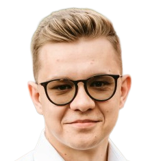
Benedikt Wiestler
is Professor for AI for Image-Guided Diagnosis and Therapy at TU Munich.
His research bridges the gap between medicine and computer science towards data-driven, personalized medicine for diagnosis and therapy. His research focuses on developing innovative computational analysis methods to extract actionable biomarkers for clinical decision-making from heterogeneous, multi-modal medical data. Translating these advancements into clinical application is a core motivation for his work.
Team members @MCML
PostDocs
PhD Students
Recent News @MCML
Publications @MCML
2026
[46]

J. Li • C. Liu • W. Bai • M. Liu • R. Arcucci • C. I. Bercea • J. A. Schnabel
Knowledge to Sight: Reasoning over Visual Attributes via Knowledge Decomposition for Abnormality Grounding.
WACV 2026 - IEEE/CVF Winter Conference on Applications of Computer Vision. Tucson, AZ, USA, Mar 06-10, 2026. To be published. Preprint available. arXiv GitHub
Knowledge to Sight: Reasoning over Visual Attributes via Knowledge Decomposition for Abnormality Grounding.
WACV 2026 - IEEE/CVF Winter Conference on Applications of Computer Vision. Tucson, AZ, USA, Mar 06-10, 2026. To be published. Preprint available. arXiv GitHub
[45]

A. Varma • D. Scholz • A. C. Erdur • J. C. Peeken • D. Rückert • B. Wiestler
Improving out-of-domain generalization in Multiple Sclerosis detection and segmentation using Random Convolutions.
Pattern Recognition Letters 199. Jan. 2026. DOI
Improving out-of-domain generalization in Multiple Sclerosis detection and segmentation using Random Convolutions.
Pattern Recognition Letters 199. Jan. 2026. DOI
[44]
D. Hooft • S. M. Fischer • C. I. Bercea • J. C. Peeken • J. A. Schnabel
LocBAM: Advancing 3D Patch-Based Image Segmentation by Integrating Location Contex.
Preprint (Jan. 2026). arXiv GitHub
LocBAM: Advancing 3D Patch-Based Image Segmentation by Integrating Location Contex.
Preprint (Jan. 2026). arXiv GitHub
[43]
B. Schaper • M. Di Folco • B. Kainz • J. A. Schnabel • C. I. Bercea
Measuring and Aligning Abstraction in Vision-Language Models with Medical Taxonomies.
Preprint (Jan. 2026). arXiv
Measuring and Aligning Abstraction in Vision-Language Models with Medical Taxonomies.
Preprint (Jan. 2026). arXiv
2025
[42]
J. Weidner • I. Ezhov • M. Balcerak • B. Menze • D. Rückert • B. Wiestler
Train Forwards, Optimize Backwards: Neural Surrogates for Personalized Medical Simulations.
DiffSys @EurIPS 2025 - Workshop on Differentiable Systems and Scientific Machine Learning at the European Conference on Information Processing Systems. Copenhagen, Denmark, Dec 03-05, 2025. To be published. Preprint available. URL
[41]
B. Jian • J. Pan • R. Jena • M. Ghahremani • H. B. Li • D. Rückert • C. Wachinger • B. Wiestler
Disentangling Progress in Medical Image Registration: Beyond Trend-Driven Architectures towards Domain-Specific Strategies.
Preprint (Dec. 2025). arXiv GitHub
Disentangling Progress in Medical Image Registration: Beyond Trend-Driven Architectures towards Domain-Specific Strategies.
Preprint (Dec. 2025). arXiv GitHub
[40]
J. McGinnis • F. A. Hölzl • S. Shit • F. Bieder • P. Friedrich • M. Mühlau • B. Menze • D. Rückert • B. Wiestler
Optimizing Rank for High-Fidelity Implicit Neural Representations.
Preprint (Dec. 2025). arXiv GitHub
Optimizing Rank for High-Fidelity Implicit Neural Representations.
Preprint (Dec. 2025). arXiv GitHub
[39]

C. I. Bercea • J. Li • P. Raffler • E. O. Riedel • L. Schmitzer • A. Kurz • F. Bitzer • P. Roßmüller • J. Canisius • M. L. Beyrle • C. Liu • W. Bai • B. Kainz • J. A. Schnabel • B. Wiestler
NOVA: A Benchmark for Rare Anomaly Localization and Clinical Reasoning in Brain MRI.
NeurIPS 2025 - 39th Conference on Neural Information Processing Systems. San Diego, CA, USA, Nov 30-Dec 07, 2025. To be published. Preprint available. URL
NOVA: A Benchmark for Rare Anomaly Localization and Clinical Reasoning in Brain MRI.
NeurIPS 2025 - 39th Conference on Neural Information Processing Systems. San Diego, CA, USA, Nov 30-Dec 07, 2025. To be published. Preprint available. URL
[38]
J. Kiechle • S. M. Fischer • D. M. Lang • C. I. Bercea • M. J. Nyflot • L. Felsner • J. A. Schnabel • J. C. Peeken
TomoGraphView: 3D Medical Image Classification with Omnidirectional Slice Representations and Graph Neural Networks.
Preprint (Nov. 2025). arXiv GitHub
TomoGraphView: 3D Medical Image Classification with Omnidirectional Slice Representations and Graph Neural Networks.
Preprint (Nov. 2025). arXiv GitHub
[37]
H. Y. Kim • J. Li • A. B. Solana • C. M. Pirkl • B. Wiestler • J. A. Schnabel • C. I. Bercea
Learning to reason about rare diseases through retrieval-augmented agents.
Preprint (Nov. 2025). arXiv
Learning to reason about rare diseases through retrieval-augmented agents.
Preprint (Nov. 2025). arXiv
[36]
N. Li • M. Ghahremani • B. Jian • P. T. Cervera • B. Wiestler • M. Makowski • C. Wachinger
Cross-Domain Semi-Supervised Organ Detection.
Preprint (Nov. 2025). URL
Cross-Domain Semi-Supervised Organ Detection.
Preprint (Nov. 2025). URL
[35]
L. Lux • A. H. Berger • M. R. Tricas • R. Rosen • A. E. Fayed • S. Sivaprasada • L. Kreitner • J. Weidner • M. J. Menten • D. Rückert • J. C. Paetzold
Interpretable Retinal Disease Prediction Using Biology-Informed Heterogeneous Graph Representations.
Preprint (Nov. 2025). arXiv
Interpretable Retinal Disease Prediction Using Biology-Informed Heterogeneous Graph Representations.
Preprint (Nov. 2025). arXiv
[34]
N. Stolt-Ansó • M. Dannecker • S. Jia • J. McGinnis • D. Rückert
NISF++: Geometrically-grounded implicit representations of 3D+time cardiac function from 2D short- and long-axis MR views.
Preprint (Nov. 2025). URL
NISF++: Geometrically-grounded implicit representations of 3D+time cardiac function from 2D short- and long-axis MR views.
Preprint (Nov. 2025). URL
[33]
A. Varma • J. Weidner • L. Lux • C. I. Bercea • M. Mühlau • J. Kirschke • B. Wiestler • D. Rückert
SegMaST: Mamba-based Spatio-Temporal Modeling to Improve Longitudinal Detection and Segmentation.
Preprint (Nov. 2025). URL
SegMaST: Mamba-based Spatio-Temporal Modeling to Improve Longitudinal Detection and Segmentation.
Preprint (Nov. 2025). URL
[32]
J. Weidner • L. Zimmer • I. Ezhov • M. Balcerak • B. Menze • D. Rückert • B. Wiestler
Brain Tumor Growth Inversion via Differentiable Neural Surrogates.
Preprint (Nov. 2025). URL
Brain Tumor Growth Inversion via Differentiable Neural Surrogates.
Preprint (Nov. 2025). URL
[31]

C. Saueressig • C. Delbridge • D. Scholz • A. Kazemi • M. Z. Khan • M. Metz • B. Meyer • M. Mitsdoerffer • P. J. Schüffler • B. Wiestler
From histology to diagnosis: Leveraging pathology foundation models for glioma classification.
Computers in Biology and Medicine 197.Part A. Oct. 2025. DOI
From histology to diagnosis: Leveraging pathology foundation models for glioma classification.
Computers in Biology and Medicine 197.Part A. Oct. 2025. DOI
[30]
V. Biller • L. Zimmer • A. C. Erdur • S. Nagar • D. Rückert • N. Bubeck • J. Weidner
A Biophysically-Conditioned Generative Framework for 3D Brain Tumor MRI Synthesis.
Preprint (Oct. 2025). arXiv GitHub
A Biophysically-Conditioned Generative Framework for 3D Brain Tumor MRI Synthesis.
Preprint (Oct. 2025). arXiv GitHub
[29]
A. C. Erdur • D. Scholz • J. A. Buchner • D. Bernhardt • S. E. Combs • B. Wiestler • D. Rückert • J. C. Peeken
Independent Benchmarking of Prompt-Based Medical Segmentation Models.
Preprint (Oct. 2025). DOI
Independent Benchmarking of Prompt-Based Medical Segmentation Models.
Preprint (Oct. 2025). DOI
[28]
J. Weidner • I. Ezhov • M. Balcerak • A. Datchev • L. Zimmer • D. Rückert • B. Menze • B. Wiestler
From Fiber Tracts to Tumor Spread: Biophysical Modeling of Butterfly Glioma Growth Using Diffusion Tensor Imaging.
CDMRI @MICCAI 2025 - Workshop on Computational Diffusion MRI at 28th International Conference on Medical Image Computing and Computer Assisted Intervention. Daejeon, Republic of Korea, Sep 23-27, 2025. DOI
[27]
M. Hartenberger • H. Ayaz • F. Ozlugedik • C. Caredda • L. Giannoni • F. Lange • L. Lux • J. Weidner • A. Berger • F. Kofler • M. J. Menten • B. Montcel • I. Tachtsidis • D. Rückert • I. Ezhov
Redefining spectral unmixing for in-vivo brain tissue analysis from hyperspectral imaging.
CMMCA @MICCAI 2025 - Workshop on Computational Mathematics Modeling in Cancer Analysis at 28th International Conference on Medical Image Computing and Computer Assisted Intervention. Daejeon, Republic of Korea, Sep 23-27, 2025. DOI
[26]
J. Weidner • M. Balcerak • I. Ezhov • A. Datchev • L. Lux • L. Zimmer • D. Rückert • B. Menze • B. Wiestler
A Lightweight Optimization Framework for Estimating 3D Brain Tumor Infiltration.
CMMCA @MICCAI 2025 - Workshop on Computational Mathematics Modeling in Cancer Analysis at 28th International Conference on Medical Image Computing and Computer Assisted Intervention. Daejeon, Republic of Korea, Sep 23-27, 2025. DOI
[25]

D. Scholz • A. C. Erdur • V. Ehm • A. Meyer-Baese • J. C. Peeken • D. Rückert • B. Wiestler
MM-DINOv2: Adapting Foundation Models for Multi-Modal Medical Image Analysis.
MICCAI 2025 - 28th International Conference on Medical Image Computing and Computer Assisted Intervention. Daejeon, Republic of Korea, Sep 23-27, 2025. DOI
MM-DINOv2: Adapting Foundation Models for Multi-Modal Medical Image Analysis.
MICCAI 2025 - 28th International Conference on Medical Image Computing and Computer Assisted Intervention. Daejeon, Republic of Korea, Sep 23-27, 2025. DOI
[24]

D. Scholz • A. C. Erdur • R. Holland • V. Ehm • J. C. Peeken • B. Wiestler • D. Rückert
Contrastive Anatomy-Contrast Disentanglement: A Domain-General MRI Harmonization Method.
MICCAI 2025 - 28th International Conference on Medical Image Computing and Computer Assisted Intervention. Daejeon, Republic of Korea, Sep 23-27, 2025. DOI
Contrastive Anatomy-Contrast Disentanglement: A Domain-General MRI Harmonization Method.
MICCAI 2025 - 28th International Conference on Medical Image Computing and Computer Assisted Intervention. Daejeon, Republic of Korea, Sep 23-27, 2025. DOI
[23]

C. K. Wong • A. N. Christensen • C. I. Bercea • J. A. Schnabel • M. G. Tolsgaard • A. Feragen
Influence of Classification Task and Distribution Shift Type on OOD Detection in Fetal Ultrasound.
MICCAI 2025 - 28th International Conference on Medical Image Computing and Computer Assisted Intervention. Daejeon, Republic of Korea, Sep 23-27, 2025. DOI GitHub
Influence of Classification Task and Distribution Shift Type on OOD Detection in Fetal Ultrasound.
MICCAI 2025 - 28th International Conference on Medical Image Computing and Computer Assisted Intervention. Daejeon, Republic of Korea, Sep 23-27, 2025. DOI GitHub
[22]
A. C. Erdur • C. Beischl • D. Scholz • J. Pan • B. Wiestler • D. Rückert • J. C. Peeken
MultiMAE for Brain MRIs: Robustness to Missing Inputs Using Multi-Modal Masked Autoencoder.
MLMI @MICCAI 2025 - 16th International Workshop on Machine Learning in Medical Imaging at the 28th International Conference on Medical Image Computing and Computer Assisted Intervention. Daejeon, Republic of Korea, Sep 23-27, 2025. DOI GitHub
[21]
S. J. Roughley • J. P. Müller • S. Gao • Z. Gao • M. Ligero • R. Blums • M. Crispin-Ortuzar • J. A. Schnabel • B. Kainz • C. I. Bercea • I. P. Machado
GroundingDINO for Open-Set Lesion Detection in Medical Imaging.
MSB EMERGE @MICCAI 2025 - 2nd MICCAI Student Board Emerge Workshop at the 28th International Conference on Medical Image Computing and Computer Assisted Intervention. Daejeon, Republic of Korea, Sep 23-27, 2025. To be published. Preprint available. URL
[20]

T. Wiltgen • J. McGinnis • R. Berg • C. C. Voon • O. Puonti • K. Giglhuber • C. Ganter • C. Zimmer • B. Hemmer • B. Wiestler • J. Kirschke • C. Preibisch • M. Mühlau
Towards quantitative intensity analysis of conventional T1-weighted images in multiple sclerosis.
NeuroImage 318.121395. Sep. 2025. DOI
Towards quantitative intensity analysis of conventional T1-weighted images in multiple sclerosis.
NeuroImage 318.121395. Sep. 2025. DOI
[19]
C. Liu • Y. Chen • H. Shi • J. Lu • B. Jian • J. Pan • L. Cai • J. Wang • Y. Zhang • J. Li • C. I. Bercea • C. Ouyang • C. Chen • Z. Xiong • B. Wiestler • C. Wachinger • D. Rückert • W. Bai • R. Arcucci
Does DINOv3 Set a New Medical Vision Standard?
Preprint (Sep. 2025). arXiv
Does DINOv3 Set a New Medical Vision Standard?
Preprint (Sep. 2025). arXiv
[18]
L. Zimmer • J. Weidner • M. Balcerak • F. Kofler • I. Ezhov • B. Menze • B. Wiestler
PREDICT-GBM: Platform for Robust Evaluation and Development of Individualized Computational Tumor Models in Glioblastoma.
Preprint (Sep. 2025). arXiv
PREDICT-GBM: Platform for Robust Evaluation and Development of Individualized Computational Tumor Models in Glioblastoma.
Preprint (Sep. 2025). arXiv
[17]
J. Pan • B. Jian • P. Hager • Y. Zhang • C. Liu • F. Jungmann • H. B. Li • C. You • J. Wu • J. Zhu • F. Liu • Y. Liu • N. Bubeck • C. Wachinger • C. Chen • Z. Gong • C. Ouyang • G. Kaissis • B. Wiestler • D. Rückert
Beyond Benchmarks: Dynamic, Automatic And Systematic Red-Teaming Agents For Trustworthy Medical Language Models.
Preprint (Aug. 2025). arXiv
Beyond Benchmarks: Dynamic, Automatic And Systematic Red-Teaming Agents For Trustworthy Medical Language Models.
Preprint (Aug. 2025). arXiv
[16]
C. Pellegrini • E. Özsoy • B. Busam • B. Wiestler • N. Navab • M. Keicher
RaDialog: Large Vision-Language Models for X-Ray Reporting and Dialog-Driven Assistance.
MIDL 2025 - Medical Imaging with Deep Learning. Salt Lake City, UT, USA, Jul 09-11, 2025. URL GitHub
RaDialog: Large Vision-Language Models for X-Ray Reporting and Dialog-Driven Assistance.
MIDL 2025 - Medical Imaging with Deep Learning. Salt Lake City, UT, USA, Jul 09-11, 2025. URL GitHub
[15]
L. A. Heidrich • A. Rastogi • P. Upadhya • G. Brugnara • M. Foltyn-Dumitru • B. Wiestler • P. Vollmuth
Curriculum Learning for Language-guided, Multi-modal Detection of Various Pathologies.
MIDL 2025 - Medical Imaging with Deep Learning. Salt Lake City, UT, USA, Jul 09-11, 2025. To be published. Preprint available. URL
Curriculum Learning for Language-guided, Multi-modal Detection of Various Pathologies.
MIDL 2025 - Medical Imaging with Deep Learning. Salt Lake City, UT, USA, Jul 09-11, 2025. To be published. Preprint available. URL
[14]
K. Yang • F. Musio • Y. Ma • N. Juchler • J. C. Paetzold • R. Al-Maskari • L. Höher • H. B. Li • I. E. Hamamci • A. Sekuboyina • S. Shit • H. Huang • C. Prabhakar • E. de la Rosa • B. Wittmann • D. Waldmannstetter • F. Kofler • F. Navarro • M. J. Menten • I. Ezhov • D. Rückert • I. N. Vos • Y. M. Ruigrok • B. K. Velthuis • H. J. Kuijf • P. Shi • W. Liu • T. Ma • M. R. Rokuss • Y. Kirchhoff • F. Isensee • K. Maier-Hein • C. Zhu • H. Zhao • P. Bijlenga • J. Hämmerli • C. Wurster • L. Westphal • J. Bisschop • E. Colombo • H. Baazaoui • H.-L. Handelsmann • A. Makmur • J. Hallinan • A. Soundararajan • B. Wiestler • J. S. Kirschke • R. Wiest • E. Montagnon • L. Letourneau-Guillon • K. Oh • D. Lee • A. Hilbert • O. U. Aydin • D. Rallios • J. Rieger • S. Tanioka • A. Koch • D. Frey • A. Qayyum • M. Mazher • S. Niederer • N. Disch • J. Holzschuh • D. LaBella • F. Galati • D. Falcetta • M. A. Zuluaga • C. Lin • H. Zhao • Z. Zhang • M. Zhang • X. You • H. Zhang • G.-Z. Yang • Y. Gu • S. Ra • J. Hwang • H. Park • J. Chen • M. Wodzinski • H. Müller • N. Mansouri • F. Autrusseau • C. Yalçin • R. E. Hamadache • C. Lisazo • J. Salvi • A. Casamitjana • X. Lladó • U. M. Lal-Trehan Estrada • V. Abramova • L. Giancardo • A. Oliver • P. Casademunt • A. Galdran • M. Delucchi • J. Liu • H. Huang • Y. Cui • Z. Lin • Y. Liu • S. Zhu • T. R. Patel • A. H. Siddiqui • V. M. Tutino • M. Orouskhani • H. Wang • M. Mossa-Basha • Y. Sato • S. Hirsch • S. Wegener • B. Menze
Benchmarking the CoW with the TopCoW Challenge: Topology-Aware Anatomical Segmentation of the Circle of Willis for CTA and MRA.
Preprint (Jul. 2025). arXiv
Benchmarking the CoW with the TopCoW Challenge: Topology-Aware Anatomical Segmentation of the Circle of Willis for CTA and MRA.
Preprint (Jul. 2025). arXiv
[13]

M. Balcerak • J. Weidner • P. Karnakov • I. Ezhov • S. Litvinov • P. Koumoutsakos • T. Amiranashvili • R. Z. Zhang • J. S. Lowengrub • I. Yakushev • B. Wiestler • B. Menze
Individualizing glioma radiotherapy planning by optimization of a data and physics-informed discrete loss.
Nature Communications 16.5982. Jun. 2025. DOI
Individualizing glioma radiotherapy planning by optimization of a data and physics-informed discrete loss.
Nature Communications 16.5982. Jun. 2025. DOI
[12]
C. J. Mertens • H. Häntze • S. Ziegelmayer • J. N. Kather • D. Truhn • S. H. Kim • F. Busch • D. Weller • B. Wiestler • M. Graf • F. Bamberg • C. L. Schlett • J. B. Weiss • S. Ringhof • E. Can • J. Schulz-Menger • T. Niendorf • J. Lammert • I. Molwitz • A. Kader • A. Hering • A. Meddeb • J. Nawabi • M. B. Schulze • T. Keil • S. N. Willich • L. Krist • M. Hadamitzky • A. Hannemann • F. Bassermann • D. Rückert • T. Pischon • A. Hapfelmeier • M. R. Makowski • K. K. Bressem • L. C. Adams
Deep learning-enabled MRI phenotyping uncovers regional body composition heterogeneity and disease associations in two European population cohorts.
Preprint (Jun. 2025). DOI
Deep learning-enabled MRI phenotyping uncovers regional body composition heterogeneity and disease associations in two European population cohorts.
Preprint (Jun. 2025). DOI
[11]
V. Ruozzi • S. Matinfar • L. Schütz • B. Wiestler • A. Redaelli • E. Votta • N. Navab
BioSonix: Can Physics-based Sonification Perceptualize Tissue Deformations From Tool Interactions?
IPMI 2025 - Information Processing in Medical Imaging. Kos Island, Greece, May 25-30, 2025. DOI
BioSonix: Can Physics-based Sonification Perceptualize Tissue Deformations From Tool Interactions?
IPMI 2025 - Information Processing in Medical Imaging. Kos Island, Greece, May 25-30, 2025. DOI
[10]
J. Li • C. Liu • W. Bai • R. Arcucci • C. I. Bercea • J. A. Schnabel
Enhancing Abnormality Grounding for Vision Language Models with Knowledge Descriptions.
Preprint (Mar. 2025). arXiv GitHub
Enhancing Abnormality Grounding for Vision Language Models with Knowledge Descriptions.
Preprint (Mar. 2025). arXiv GitHub
[9]

C. I. Bercea • B. Wiestler • D. Rückert • J. A. Schnabel
Evaluating normative representation learning in generative AI for robust anomaly detection in brain imaging.
Nature Communications 16.1624. Feb. 2025. DOI GitHub
Evaluating normative representation learning in generative AI for robust anomaly detection in brain imaging.
Nature Communications 16.1624. Feb. 2025. DOI GitHub
[8]
Z. Haouari • J. Weidner • I. Ezhov • A. Varma • D. Rückert • B. Menze • B. Wiestler
Efficient Deep Learning-based Forward Solvers for Brain Tumor Growth Models.
Preprint (Jan. 2025). arXiv
Efficient Deep Learning-based Forward Solvers for Brain Tumor Growth Models.
Preprint (Jan. 2025). arXiv
[7]
B. Jian • J. Pan • Y. Li • F. Bongratz • R. Li • D. Rückert • B. Wiestler • C. Wachinger
TimeFlow: Longitudinal Brain Image Registration and Aging Progression Analysis.
Preprint (Jan. 2025). arXiv
TimeFlow: Longitudinal Brain Image Registration and Aging Progression Analysis.
Preprint (Jan. 2025). arXiv
2024
[6]
J. Li • S. H. Kim • P. Müller • L. Felsner • D. Rückert • B. Wiestler • J. A. Schnabel • C. I. Bercea
Language Models Meet Anomaly Detection for Better Interpretability and Generalizability.
MMMI @MICCAI 2024 - 5th International Workshop on Multiscale Multimodal Medical Imaging at the 27th International Conference on Medical Image Computing and Computer Assisted Intervention. Marrakesh, Morocco, Oct 06-10, 2024. DOI GitHub
[5]
B. Jian • J. Pan • M. Ghahremani • D. Rückert • C. Wachinger • B. Wiestler
Mamba? Catch The Hype Or Rethink What Really Helps for Image Registration.
WBIR @MICCAI 2024 - 11th International Workshop on Biomedical Image Registration at the 27th International Conference on Medical Image Computing and Computer Assisted Intervention. Marrakesh, Morocco, Oct 06-10, 2024. DOI
Mamba? Catch The Hype Or Rethink What Really Helps for Image Registration.
WBIR @MICCAI 2024 - 11th International Workshop on Biomedical Image Registration at the 27th International Conference on Medical Image Computing and Computer Assisted Intervention. Marrakesh, Morocco, Oct 06-10, 2024. DOI
[4]

A. C. Erdur • D. Rusche • D. Scholz • J. Kiechle • S. M. Fischer • Ó. Llorián-Salvador • J. A. Buchner • M. Q. Nguyen • L. Etzel • J. Weidner • M.-C. Metz • B. Wiestler • J. A. Schnabel • D. Rückert • S. E. Combs • J. C. Peeken
Deep learning for autosegmentation for radiotherapy treatment planning: State-of-the-art and novel perspectives.
Strahlentherapie und Onkologie 201. Aug. 2024. DOI GitHub
Deep learning for autosegmentation for radiotherapy treatment planning: State-of-the-art and novel perspectives.
Strahlentherapie und Onkologie 201. Aug. 2024. DOI GitHub
2023
[3]
D. Scholz • B. Wiestler • D. Rückert • M. J. Menten
Metrics to Quantify Global Consistency in Synthetic Medical Images.
DGM4 @MICCAI 2023 - 3rd International Workshop on Deep Generative Models at the 26th International Conference on Medical Image Computing and Computer Assisted Intervention. Vancouver, Canada, Oct 08-12, 2023. DOI
Metrics to Quantify Global Consistency in Synthetic Medical Images.
DGM4 @MICCAI 2023 - 3rd International Workshop on Deep Generative Models at the 26th International Conference on Medical Image Computing and Computer Assisted Intervention. Vancouver, Canada, Oct 08-12, 2023. DOI
[2]

N. Stolt-Ansó • J. McGinnis • J. Pan • K. Hammernik • D. Rückert
NISF: Neural implicit segmentation functions.
MICCAI 2023 - 26th International Conference on Medical Image Computing and Computer Assisted Intervention. Vancouver, Canada, Oct 08-12, 2023. DOI
NISF: Neural implicit segmentation functions.
MICCAI 2023 - 26th International Conference on Medical Image Computing and Computer Assisted Intervention. Vancouver, Canada, Oct 08-12, 2023. DOI
2022
[1]
P. Engstler • M. Keicher • D. Schinz • K. Mach • A. S. Gersing • S. C. Foreman • S. S. Goller • J. Weissinger • J. Rischewski • A.-S. Dietrich • B. Wiestler • J. S. Kirschke • A. Khakzar • N. Navab
Interpretable Vertebral Fracture Diagnosis.
iMIMIC @MICCAI 2022 - Workshop on Interpretability of Machine Intelligence in Medical Image Computing at the 25th International Conference on Medical Image Computing and Computer Assisted Intervention. Singapore, Sep 18-22, 2022. DOI GitHub
Interpretable Vertebral Fracture Diagnosis.
iMIMIC @MICCAI 2022 - Workshop on Interpretability of Machine Intelligence in Medical Image Computing at the 25th International Conference on Medical Image Computing and Computer Assisted Intervention. Singapore, Sep 18-22, 2022. DOI GitHub
©all images: LMU | TUM


